

Perioperative visual loss in ocular and nonocular surgery
- PMID: 20596508
- PMCID: PMC2893763
- DOI: 10.2147/opth.s9262
Perioperative visual loss in ocular and nonocular surgery
Abstract
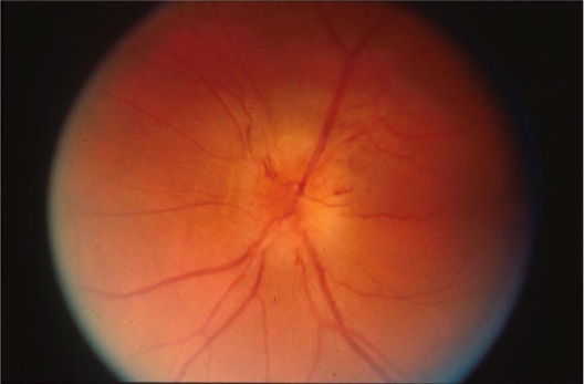
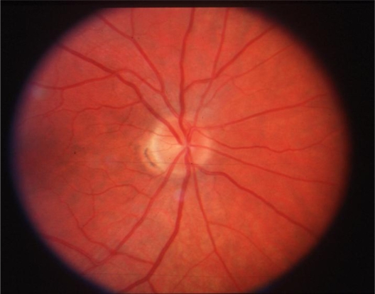
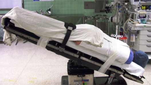
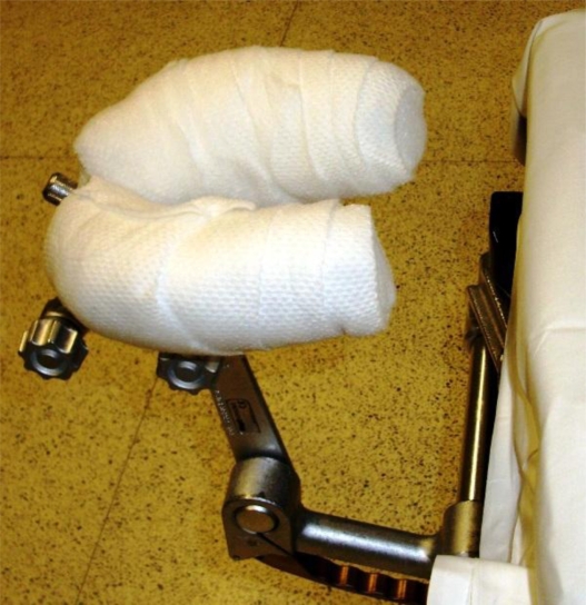
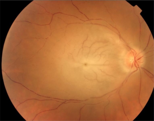
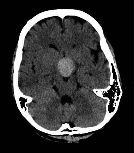
Incidence estimates for perioperative vision loss (POVL) after nonocular surgery range from 0.013% for all surgeries up to 0.2% following spine surgery. The most common neuro-ophthalmologic causes of POVL are the ischemic optic neuropathies (ION), either anterior (AION) or posterior (PION). We identified 111 case reports of AION following nonocular surgery in the literature, with most occurring after cardiac surgery, and 165 case reports of PION following nonocular surgery, with most occurring after spine surgery or radical neck dissection. There were an additional 526 cases of ION that did not specify if the diagnosis was AION or PION. We also identified 933 case reports of central retinal artery occlusion (CRAO), 33 cases of pituitary apoplexy, and 245 cases of cortical blindness following nonocular surgery. The incidence of POVL following ocular surgery appears to be much lower than that seen following nonocular surgery. We identified five cases in the literature of direct optic nerve trauma, 47 cases of AION, and five cases of PION following ocular surgery. The specific pathogenesis and risk factors underlying these neuro-ophthalmic complications remain unknown, and physicians should be alert to the potential for loss of vision in the postoperative period.
Keywords: nonocular surgery; ocular surgery; perioperative; postoperative; vision loss.
Figures
References
-
- Warner ME, Warner MA, Garrity JA, MacKenzie RA, Warner DO. The frequency of perioperative vision loss. Anesth Analg. 2001;93(6):1417–1421. - PubMed
-
- Roth S, Thisted RA, Erickson JP, Black S, Schreider BD. Eye injuries after nonocular surgery. A study of 60,965 anesthetics from 1988 to 1992. Anesthesiology. 1996;85(5):1020–1027. - PubMed
-
- Shen Y, Drum M, Roth S. The prevalence of perioperative visual loss in the United States: a 10-year study from 1996 to 2005 of spinal, orthopedic, cardiac, and general surgery. Anesth Analg. 2009;109(5):1534–1545. - PubMed
-
- Nuttall GA, Garrity JA, Dearani JA, Abel MD, Schroeder DR, Mullany CJ. Risk factors for ischemic optic neuropathy after cardiopulmonary bypass: a matched case/control study. Anesth Analg. 2001;93(6):1410–1416. - PubMed
-
- Sweeney PJ, Breuer AC, Selhorst JB, et al. Ischemic optic neuropathy: a complication of cardiopulmonary bypass surgery. Neurology. 1982;32(5):560–562. - PubMed
LinkOut - more resources
Full Text Sources
