. 2010 Jun 28;5(6):e11247.
doi: 10.1371/journal.pone.0011247.

MAPK-activated protein kinase 2 is required for mouse meiotic spindle assembly and kinetochore-microtubule attachment

Affiliations

MAPK-activated protein kinase 2 is required for mouse meiotic spindle assembly and kinetochore-microtubule attachment

Ju Yuan et al. PLoS One. .

Abstract

MAPK-activated protein kinase 2 (MK2), a direct substrate of p38 MAPK, plays key roles in multiple physiological functions in mitosis. Here, we show for the first time the unique distribution pattern of MK2 in meiosis. Phospho-MK2 was localized on bipolar spindle minus ends and along the interstitial axes of homologous chromosomes extending over centromere regions and arm regions at metaphase of first meiosis (MI stage) in mouse oocytes. At metaphase of second meiosis (MII stage), p-MK2 was localized on the bipolar spindle minus ends and at the inner centromere region of sister chromatids as dots. Knockdown or inhibition of MK2 resulted in spindle defects. Spindles were surrounded by irregular nondisjunction chromosomes, which were arranged in an amphitelic or syntelic/monotelic manner, or chromosomes detached from the spindles. Kinetochore-microtubule attachments were impaired in MK2-deficient oocytes because spindle microtubules became unstable in response to cold treatment. In addition, homologous chromosome segregation and meiosis progression were inhibited in these oocytes. Our data suggest that MK2 may be essential for functional meiotic bipolar spindle formation, chromosome segregation and proper kinetochore-microtubule attachments.


Conflict of interest statement

Competing Interests: The authors have declared that no competing interests exist.

Figures

Figure 1
Figure 1. Expression and subcellular localization of p-MK2 during mouse oocyte meiotic maturation.
Samples were collected after oocytes had been cultured for 0, 4, 8, 9.5 and 12 h, corresponding to GV, Pro-MI, MI, AI to TI and MII stages, respectively. Proteins from a total of 200 oocytes were loaded for each sample. (B) Oocytes at various stages were double stained with antibodies against p-MK2 (green) and with propidium iodide (PI, red). GV, oocytes at germinal vesicle stage; Pro-MI, oocytes at first prometaphase; MI, oocytes at first metaphase; AI, oocytes at anaphase; TI, oocytes at telophase; MII, oocytes at second metaphase. Bar 20 µm. (C) Metaphase I and metaphase II oocytes were fixed and double labeled with rabbit p-MK2 antibody (red) and human Crest antibody (green). Each sample was counterstained with Hoechst 33258 to visualize DNA (blue). Bar 20 µm. (a) model chart for localization of p-MK2, Crest and chromosomes at MI stage oocytes. (b) model chart for localization of p-MK2, Crest and chromosomes at MII stage oocytes. (D) Oocytes cultured for 12h (MII) were fixed and stained for γ- tubulin (green), p-MK2 (red) and DNA (blue) as visualized with Hoechst 33258 staining. Bar 20 µm.
Figure 2
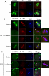
Figure 2. Localization of p-MK2 in mouse oocytes treated with taxol and nocodazole.
(A) Oocytes cultured for 8.5 h (MI) and 12 h (MII) were incubated in M16 medium containing 10 µM taxol for 45 min and then double stained with antibodies against p-MK2 (red), α-tubulin (green), and DNA (blue). (B) Oocytes at the metaphase I stage were incubated in M16 medium containing 20 µg/ml nocodazole for 10 min (b,c) and then washed thoroughly and cultured in fresh M16 mediun for 30 min (d). Control groups were treated with DMSO (a). Then oocytes were fixed and double stained with antibodies against p-MK2 (red), α-tubulin (green), and stained for DNA (blue). Bar 20 µm. (C) Oocytes at the metaphase II stage were incubated in M16 medium containing 20 µg/ml nocodazole for 10 min (b) and then washed thoroughly and cultured in fresh M16 medium for 30 min (c). Control group was treated with DMSO(a). Then oocytes were fixed and double stained with antibodies against p-MK2 (red), α-tubulin (green), and DNA (blue). Bar 20 µm.
Figure 3
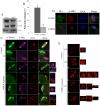
Figure 3. Depletion of MK2 by morpholino injection induced homologous chromosome misalignment and bipolar spindle assembly defects.
(A) Samples from control and MK2 MO injection groups were collected to test the efficiency of MK2 depletion. A total of 200 oocytes were injected with control or MK2 morpholino and cultured for 6 h after arrest at the GV stage for 24 h in M2-containing 2.5 µM milrinone. (B) Percentage of polar body extrusion in the MK2 morpholino injected group (n = 196) and control morpholino injected group (n = 137). Data are presented as mean ± SE. Different superscripts indicate statistical difference (p<0.05). (C) Immunostaining of oocytes injected with control and MK2 morpholino. After injection, oocytes were incubated for 12 h after arrest at the GV stage for 24 h, followed by immunostaining with α-tubulin antibody (green), p-MK2 (purple) and PI labeling for DNA (red). Bar 20 µm. (D) The above oocytes were immunostained with γ- tubulin (green), p-MK2 (red) and labeled for DNA (blue). Bar 20 µm. (E) The above oocytes were immunostained with Plk1 (green) and labeled with PI (red). Bar 20 µm.
Figure 4
Figure 4. MK2 inhibitor CMPD1 treatment impairs spindle organization and chromosome alignment in mouse oocytes.
(A) Samples from control and CMPD1 groups were collected to test the efficiency of the MK2 inhibitor. CMPD1, 200 oocytes were cultured in M16 medium with 30 µM CMPD1 for 10 h; Control, 200 oocytes were cultured in M16 medium with DMSO for 10h. (B) The rate of oocytes with first polar body in the control group (n = 267) and CMPD1-treated group (n = 318). Data are presented as mean percentage (mean ± SEM) of at least three independent experiments. Different superscripts indicate statistical difference (p<0.05). (C) Spindle morphologies and chromosome alignment in oocytes cultured with DMSO or MK2 inhibitor CMPD1. GV oocytes were cultured in M16 medium with DMSO or with 30 µM CMPD1 for 12 h and then stained for p-MK2 (purple), α-tubulin (green) and DNA (red). Bar 20 µm. (D) GV oocytes were cultured in M16 medium with DMSO or with 30 µM CMPD1 for 8.5 h and then stained for p-MK2 (red), Plk1 (green) and DNA (blue). Bar 20 µm. (E) Chromosome spreading was performed in oocytes that had been cultured for 8.5 h or 12 h of DMSO treatment (MI) (MII) or for 12 h of CMPD1 treatment. Representative images of each sample are shown. Bar 10 µm.
Figure 5
Figure 5. CMPD1 treatment leads to metaphase II spindle assembly defects, misaligned chromosomes and aneuploidy.
(A) The rate of oocytes with first polar body in the control group and CMPD1-treated group. Control group (−−): oocytes cultured with DMSO for 15 h (n = 100); CMPD1-treated group (−+): oocytes cultured with DMSO for 10 h, and then with 30 µM CMPD1 for 5 h (n = 265). Data are presented as mean percentage (mean ± SEM) of at least three independent experiments. Different superscripts indicate statistical difference (p<0.05). (B) Spindle morphologies and chromosome alignment in the oocytes first cultured with DMSO for 10 h, and then with MK2 inhibitor CMPD1 for 5 h; then stained for p-MK2 (purple), α-tubulin (green) and DNA (red). Control group oocytes were cultured with DMSO for 15 h. Scale bar, 20 µm. (C) GV oocytes were treated as in (B), and then stained for p-MK2 (red), Plk1 (green) and DNA (blue). Bar 20 µm. (D) Chromosome spreading was performed in oocytes that had been treated as (B). Representative images for each sample are shown. Bar 10 µm.
Figure 6
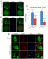
Figure 6. MK2 depletion interferes with correct microtubule–kinetochore attachment.
(A) After culture for 12 h in M16 medium at 37°C, oocytes injected with control or MK2 morpholino were quickly transferred to ice-cold medium at 4°C for 20 min. These oocytes were fixed and stained for α-tubulin (green) and DNA (blue). Scale bar, 20 µm. (B) After 8.5 h of culture in M16 medium with DMSO or CMPD1 at 37°C, oocytes were quickly transferred to ice-cold medium at 4°C for 20 min. These oocytes were fixed and stained for α-tubulin (green) and DNA (blue). Scale bar, 10 µm. (C) Percentages of oocytes with stable K-MTs after cold treatment as in (A,B). (D) Oocytes cultured in medium with DMSO or CMPD1 for 8.5 h were incubated in M16 medium containing 10 µM taxol for 45 min and then double stained with antibodies against p-MK2 (red), α-tubulin (green), and DNA label (blue). Bar 20 µm. White arrowhead indicates homologous chromosome attachment to microtubules.

Similar articles

Cited by

References

    1. Nasmyth K. Disseminating the genome: joining, resolving, and separating sister chromatids during mitosis and meiosis. Annu Rev Genet. 2001;35:673–745. - PubMed
    1. Marston AL, Amon A. Meiosis: cell-cycle controls shuffle and deal. Nat Rev Mol Cell Biol. 2004;5:983–997. - PubMed
    1. Kline-Smith SL, Walczak CE. Mitotic spindle assembly and chromosome segregation: refocusing on microtubule dynamics. Mol Cell. 2004;15:317–327. - PubMed
    1. Piehl M, Tulu US, Wadsworth P, Cassimeris L. Centrosome maturation: measurement of microtubule nucleation throughout the cell cycle by using GFP-tagged EB1. Proc Natl Acad Sci U S A. 2004;101:1584–1588. - PMC - PubMed
    1. Tong C, Fan HY, Lian L, Li SW, Chen DY, et al. Polo-like kinase-1 is a pivotal regulator of microtubule assembly during mouse oocyte meiotic maturation, fertilization, and early embryonic mitosis. Biol Reprod. 2002;67:546–554. - PubMed

Publication types

LinkOut - more resources