

Rapid and sensitive detection of Yersinia pestis using amplification of plague diagnostic bacteriophages monitored by real-time PCR
- PMID: 20596528
- PMCID: PMC2893161
- DOI: 10.1371/journal.pone.0011337
Rapid and sensitive detection of Yersinia pestis using amplification of plague diagnostic bacteriophages monitored by real-time PCR
Abstract
Background: Yersinia pestis, the agent of plague, has caused many millions of human deaths and still poses a serious threat to global public health. Timely and reliable detection of such a dangerous pathogen is of critical importance. Lysis by specific bacteriophages remains an essential method of Y. pestis detection and plague diagnostics.
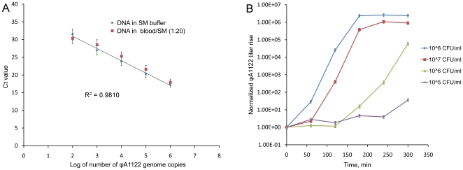
Methodology/principal findings: The objective of this work was to develop an alternative to conventional phage lysis tests--a rapid and highly sensitive method of indirect detection of live Y. pestis cells based on quantitative real-time PCR (qPCR) monitoring of amplification of reporter Y. pestis-specific bacteriophages. Plague diagnostic phages phiA1122 and L-413C were shown to be highly effective diagnostic tools for the detection and identification of Y. pestis by using qPCR with primers specific for phage DNA. The template DNA extraction step that usually precedes qPCR was omitted. phiA1122-specific qPCR enabled the detection of an initial bacterial concentration of 10(3) CFU/ml (equivalent to as few as one Y. pestis cell per 1-microl sample) in four hours. L-413C-mediated detection of Y. pestis was less sensitive (up to 100 bacteria per sample) but more specific, and thus we propose parallel qPCR for the two phages as a rapid and reliable method of Y. pestis identification. Importantly, phiA1122 propagated in simulated clinical blood specimens containing EDTA and its titer rise was detected by both a standard plating test and qPCR.
Conclusions/significance: Thus, we developed a novel assay for detection and identification of Y. pestis using amplification of specific phages monitored by qPCR. The method is simple, rapid, highly sensitive, and specific and allows the detection of only live bacteria.
Conflict of interest statement
Figures
Similar articles
-
'Bioluminescent' reporter phage for the detection of Category A bacterial pathogens.J Vis Exp. 2011 Jul 8;(53):e2740. doi: 10.3791/2740. J Vis Exp. 2011. PMID: 21775956 Free PMC article.
-
Development of a PCR-lateral flow assay for rapid detection of Yersinia pestis, the causative agent of plague.Acta Trop. 2021 Aug;220:105958. doi: 10.1016/j.actatropica.2021.105958. Epub 2021 May 15. Acta Trop. 2021. PMID: 34004173
-
Development and evaluation of a multi-target droplet digital PCR assay for highly sensitive and specific detection of Yersinia pestis.PLoS Negl Trop Dis. 2024 May 3;18(5):e0012167. doi: 10.1371/journal.pntd.0012167. eCollection 2024 May. PLoS Negl Trop Dis. 2024. PMID: 38701065 Free PMC article.
-
Bacteriophages of Yersinia pestis.Adv Exp Med Biol. 2016;918:361-375. doi: 10.1007/978-94-024-0890-4_13. Adv Exp Med Biol. 2016. PMID: 27722870 Review.
-
[Methods of diagnosis and differentiation of plague pathogen: approaches to detection of atypical strains of Yersinia pestis by molecular biology. Part I].Mol Gen Mikrobiol Virusol. 2006;(1):3-6. Mol Gen Mikrobiol Virusol. 2006. PMID: 16512602 Review. Russian.
Cited by
-
Bacteriophage-resistant mutants in Yersinia pestis: identification of phage receptors and attenuation for mice.PLoS One. 2011;6(9):e25486. doi: 10.1371/journal.pone.0025486. Epub 2011 Sep 28. PLoS One. 2011. PMID: 21980477 Free PMC article.
-
Highly Sensitive Bacteriophage-Based Detection of Brucella abortus in Mixed Culture and Spiked Blood.Viruses. 2017 Jun 10;9(6):144. doi: 10.3390/v9060144. Viruses. 2017. PMID: 28604602 Free PMC article.
-
'Bioluminescent' reporter phage for the detection of Category A bacterial pathogens.J Vis Exp. 2011 Jul 8;(53):e2740. doi: 10.3791/2740. J Vis Exp. 2011. PMID: 21775956 Free PMC article.
-
Identification and Characterization of vB_PreP_EPr2, a Lytic Bacteriophage of Pan-Drug Resistant Providencia rettgeri.Viruses. 2022 Mar 29;14(4):708. doi: 10.3390/v14040708. Viruses. 2022. PMID: 35458437 Free PMC article.
-
Yersinia pestis detection by loop-mediated isothermal amplification combined with magnetic bead capture of DNA.Braz J Microbiol. 2018 Jan-Mar;49(1):128-137. doi: 10.1016/j.bjm.2017.03.014. Epub 2017 Aug 26. Braz J Microbiol. 2018. PMID: 28887007 Free PMC article.
References
-
- Gage KL, Kosoy MY. Natural history of plague: perspectives from more than a century of research. Annu Rev Entomol. 2005;50:505–528. - PubMed
-
- Butler T. Plague into the 21st Century. Clin Infect Dis. 2009;49:736–742. - PubMed
-
- Inglesby TV, Dennis DT, Henderson DA, Bartlett JG, Ascher MS, et al. Plague as a biological weapon: medical and public health management. Working Group on Civilian Biodefense. JAMA. 2000;283:2281–2290. - PubMed
Publication types
MeSH terms
Substances
LinkOut - more resources
Full Text Sources
Other Literature Sources
Medical
Molecular Biology Databases
