

Syphilis: the renaissance of an old disease with oral implications
- PMID: 20596972
- PMCID: PMC2811633
- DOI: 10.1007/s12105-009-0127-0
Syphilis: the renaissance of an old disease with oral implications
Abstract
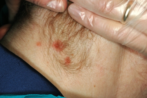
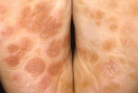
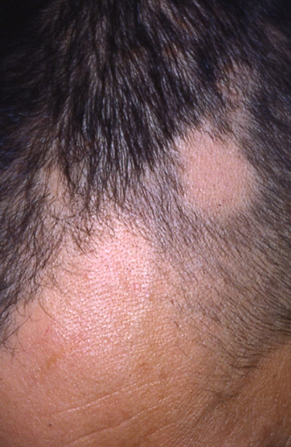
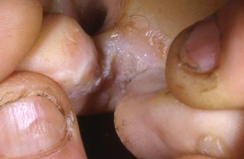
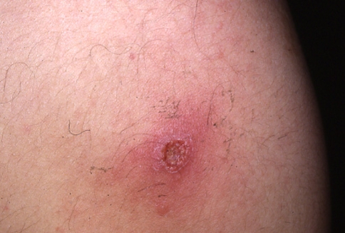
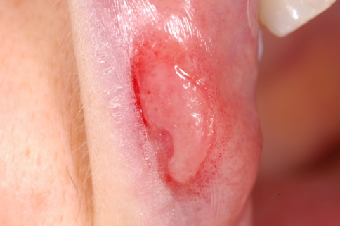
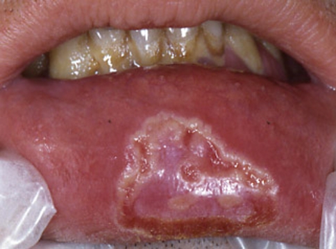
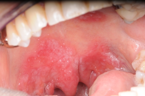
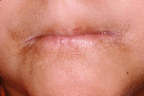
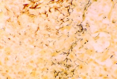
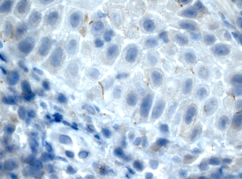
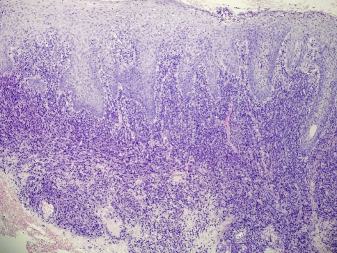
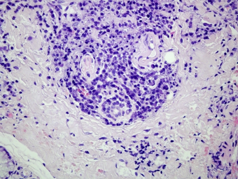
Syphilis is caused by Treponema pallidum an anaerobic filamentous spirochete. In recent years, striking outbreaks have occurred in USA, Canada, Russia, China and some areas of Central and Eastern Europe. Main epidemiology changes reflect sex industry, sexual promiscuity, decreasing use of barrier protection (i.e. condoms) due to false sense of security that nowadays sexually transmitted diseases are curable and lack of pertinent knowledge. Considering that the initial presentation of syphilis may be the oral cavity, it is of great relevance to include this disease in the differential diagnosis of unusual oral ulcerations and white patches. Primary syphilis is a highly infectious disease in which inappropriate treatment may be apparently curative while the patient remains highly infectious. It is then of pivotal importance that clinicians maintain a high clinical index of suspicion. At the present time, clinical-pathologic correlation together with serologic studies remain essential in establishing the diagnosis of syphilis.
Figures
References
-
- Hook EW, III, Marra CM. Acquired syphilis in adults. New Engl J Med. 1992;326:1060–1068. - PubMed
-
- Tramont EC. Treponema pallidum (syphilis) In: Mandell GL, Benett JF, Dolin R, editors. Principles and practice of infectious diseases. 6. Orlando, FL: Churchill Livingstone; 2005. pp. 2768–2784.
-
- Sanchez MR. Syphilis. In: Wolff K, Goldsmith LA, Katz SI, Gilchrest BA, Paller AS, Leffell DJ, editors. Fitzpatrick’s dermatology in general medicine. 7. New York: Mc Graw Hill; 2008. pp. 1955–1977.
-
- Tramont EC. Syphilis in adults: from Christopher Columbus to Sir Alexander Fleming to AIDS. Clin Infect Dis. 1995;21:1361–1369. - PubMed
Publication types
MeSH terms
LinkOut - more resources
Full Text Sources
Medical
