

What's new in the AFIP fascicle on salivary gland tumors: a few highlights from the 4th Series Atlas
- PMID: 20596976
- PMCID: PMC2811626
- DOI: 10.1007/s12105-009-0128-z
What's new in the AFIP fascicle on salivary gland tumors: a few highlights from the 4th Series Atlas
Abstract
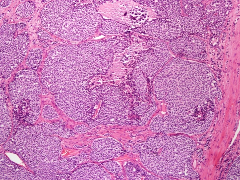
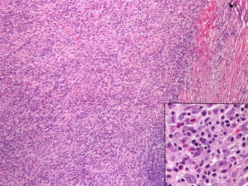
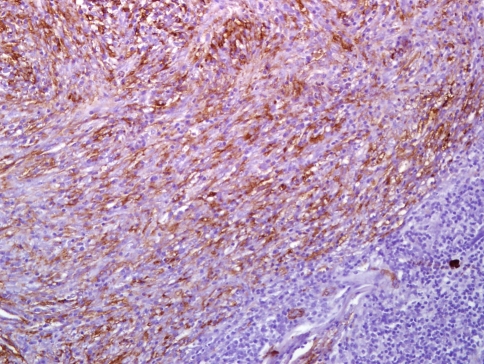
After a 12 year interval from the previous fascicle, a new fascicle on Tumors of the Salivary Glands in the new fourth series of the AFIP Atlas of Tumor Pathology was published in 2008. The data, presentation, illustrations, tables, and physical characteristics of the newest fascicle have been updated and improved. There have only been a few alterations and additions to the classification of tumors and tumor-like non-neoplastic conditions of salivary gland. Three of the most significant are discussed in this paper. Sialoblastoma has been reclassified as malignant; inflammatory pseudotumor has been reclassified as neoplastic and re-identified as inflammatory myofibroblastic tumor; and sclerosing polycystic adenosis is a new entity among tumor-like conditions.
Figures

References
-
- Ellis GL, Auclair PL. Tumors of the salivary glands. atlas of tumor pathology, 3rd series, fascicle 17. Washington DC: Armed Forces Institute of Pathology; 1996.
-
- Ellis GL, Auclair PL. Tumors of the salivary glands. AFIP atlas of tumor pathology, 4th series, fascicle 9. Silver Spring MD: ARP Press; 2008.
-
- Seifert G, Sobin LH. Histological typing of salivary gland tumours. World Health Organization international histological classification of tumours. New York: Springer; 1991.
-
- Barnes L, Eveson J, Reichart P, et al. World health organization classification of tumours. pathology and genetics of head and neck tumours. Lyon: IARC Press; 2005.
MeSH terms
LinkOut - more resources
Full Text Sources
Medical
