Diagnostically challenging epithelial odontogenic tumors: a selective review of 7 jawbone lesions
- PMID: 20596984
- PMCID: PMC2807539
- DOI: 10.1007/s12105-009-0107-4
Diagnostically challenging epithelial odontogenic tumors: a selective review of 7 jawbone lesions
Abstract
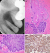
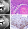
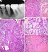
Considerable variation in the clinicopathologic presentation of epithelial odontogenic tumors can sometimes be confusing and increase the chance of misdiagnosis. Seven diagnostically challenging jawbone lesions are described. There were 2 cases of mistaken identity in our ameloblastoma file. One unicystic type, initially diagnosed and treated as a lateral periodontal cyst, showed destructive recurrence 6 years postoperatively. The other globulomaxillary lesion was managed under the erroneous diagnosis of adenomatoid odontogenic tumor and recurred 4 times over an 11-year period. This tumor was found in retrospect to be consistent with an adenoid ameloblastoma with dentinoid. The diagnosis of cystic squamous odontogenic tumor (SOT) occurring as a radicular lesion of an impacted lower third molar was one of exclusion. Of two unsuspected keratocystic odontogenic tumors, one depicted deceptive features of pericoronitis, while the other case has long been in our files with the diagnosis of globulomaxillary SOT. Two cases of primary intraosseous squamous cell carcinoma appeared benign clinically and exhibited unexpected findings; an impacted third molar began to erupt in association with the growth of carcinoma and another periradicular carcinoma showed dentinoid formation. Cases selectively reviewed in this article present challenging problems which require clinical and radiographic correlation to avoid potential diagnostic pitfalls.
Keywords: Clinicopathologic correlation; Diagnostic pitfall; Epithelial odontogenic tumor; Jawbone.
Figures


References
-
- Slootweg PJ. Lesions of the jaws. Histopathology. 2008. doi: 10.1111/j.1365-2559.2008.03097.x. - PubMed
-
- Ide F, Obara K, Yamada H, et al. Hamartomatous proliferations of odontogenic epithelium within the jaws: a potential histogenetic source of intraosseous epithelial odontogenic tumors. J Oral Pathol Med. 2007;36:229–35. - PubMed
-
- Ide F, Mishima K, Yamada H, et al. Unsuspected small ameloblastoma in the alveolar bone: a collaborative study of 14 cases with discussion of their cellular sources. J Oral Pathol Med. 2008;37:221–7. - PubMed
-
- Abaza NA, Gold L, Lally E. Granular cell odontogenic cyst: a unicytic ameloblastoma with late recurrence as follicular ameloblastoma. J Oral Maxillofac Surg. 1989;47:168–75. - PubMed
Publication types
MeSH terms
LinkOut - more resources
Full Text Sources

