

An update on grading of salivary gland carcinomas
- PMID: 20596994
- PMCID: PMC2807532
- DOI: 10.1007/s12105-009-0102-9
An update on grading of salivary gland carcinomas
Abstract
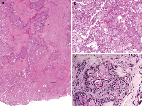
Histologic grade is a significant predictor of outcome in salivary gland carcinomas. However, the sheer variety of tumor type and the rarity of these tumors pose challenges to devising highly predictive grading schemes. As our knowledge base has evolved, it is clear that carcinoma ex pleomorphic adenoma is not automatically a high grade tumor as is traditionally suggested. These tumors should be further qualified as to type/grade of carcinoma and extent, since intracapsular and minimally invasive carcinomas ex pleomorphic adenoma behave favorably. The two carcinoma types for which grading schemes are common include adenoid cystic carcinoma and mucoepidermoid carcinoma. Adenoid cystic carcinomas are graded based solely on pattern with solid components portending a worse prognosis. Occasionally, adenoid cystic carcinomas may undergo transformation to pleomorphic high grade carcinomas. This feature confers a high propensity for lymph node metastasis and should thus be reported to alert the clinical team. Mucoepidermoid carcinomas are graded in a three tier fashion based on a constellation of features including cystic component, border, mitoses, anaplasia, and perineural invasion among others. All grading schemes are somewhat cumbersome, intimidating and occasionally ambiguous, but evidence suggests that using a scheme consistently shows greater reproducibility than using an intuitive approach. The intermediate grade category demonstrates the most variability between grading systems and thus the most controversy in management. In the AFIP system intermediate grade tumors cluster with high grade tumors, while in the Brandwein system, they cluster with low grade tumors.
Keywords: Adenoid cystic carcinoma; Carcinoma ex pleomorphic adenoma; Grading; High grade transformation; Mucoepidermoid carcinoma; Salivary carcinoma.
Figures
References
-
- Eveson JW, Auclair PL, Gnepp DR, et al. Tumors of the salivary glands: introduction. In: Barnes EL, Eveson JW, Reichart P, Sidransky D, editors. World Health Organization classification of tumours: pathology & genetics. Head and neck tumours. Lyon: IARCPress; 2005. p. 221–2.
-
- Myers EN, Ferris RL, editors. Salivary gland disorders. Berlin: Springer; 2007.
Publication types
MeSH terms
LinkOut - more resources
Full Text Sources
Medical
Research Materials
