Review
. 2009 Mar;3(1):86-93.
doi: 10.1007/s12105-009-0109-2. Epub 2009 Feb 28.

Update to the College of American Pathologists reporting on thyroid carcinomas

Affiliations
Review

Update to the College of American Pathologists reporting on thyroid carcinomas

Ronald Ghossein. Head Neck Pathol. 2009 Mar.

Abstract

Background: The reporting of thyroid carcinomas follows the recommendations of the College of American Pathologists (CAP) protocols and includes papillary carcinoma, follicular carcinoma, anaplastic carcinoma and medullary carcinoma. Despite past and recent efforts, there are a number of controversial issues in the classification and diagnosis of thyroid carcinomas (TC) that, potentially impact on therapy and prognosis of patients with TC.

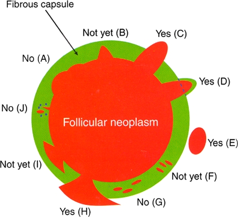
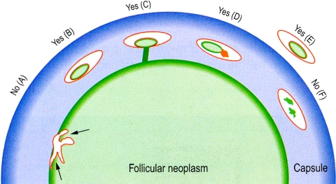
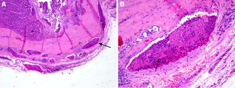
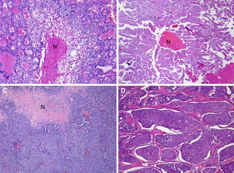
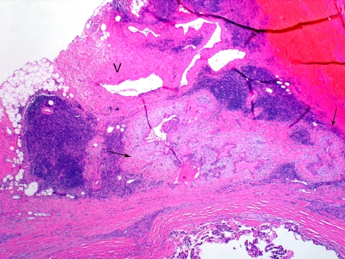
Discussion: The most updated version of the CAP thyroid cancer protocol incorporates recent changes in histologic classification as well as changes in the staging of thyroid cancers as per the updated American Joint Commission on Cancer staging manual. Among the more contentious issues in the pathology of thyroid carcinoma include the defining criteria for tumor invasiveness. While there are defined criteria for invasion, there is not universal agreement in what constitutes capsular invasion, angioinvasion and extrathyroidal invasion. Irrespective of the discrepant views on invasion, pathologists should report on the presence and extent (focal, widely) of capsular invasion, angioinvasion and extrathyroidal extension. These findings assist clinicians in their assessment of the recurrence risk and potential for metastatic disease. It is beyond the scope of this paper to detail the entire CAP protocol for thyroid carcinomas; rather, this paper addresses some of the more problematic issues confronting pathologists in their assessment and reporting of thyroid carcinomas.

Conclusion: The new CAP protocol for reporting of thyroid carcinomas is a step toward improving the clinical value of the histopathologic reporting of TC. Large meticulous clinico-pathologic and molecular studies with long term follow up are still needed in order to increase the impact of microscopic examination on the prognosis and management of TC.

Keywords: CAP; Capsular; Carcinoma; Extension; Extrathyroid; Invasion; Margins; Minimally invasive; Mitosis; Necrosis; Papillary microcarcinomas; Reporting; Thyroid; Vascular; Widely invasive.


Figures

Fig. 1
Fig. 1
Capsular invasion (CI): Schematic drawing for the interpretation of the presence or absence of CI. The diagram depicts a follicular neoplasm (orange) surrounded by a fibrous capsule (green). a bosselation on the inner aspect of the capsule does not represent CI; b sharp tumor bud invades into but not through the capsule suggesting invasion requiring deeper sections to exclude; c: tumor totally transgresses the capsule invading beyond the outer contour of the capsule qualifying as CI; d: tumor clothed by thin (probably new) fibrous capsule but already extending beyond an imaginary (dotted) line drawn through the outer contour of the capsule qualifying as CI; e: satellite tumor nodule with similar features (architecture, cytomorphology) to the main tumor lying outside the capsule qualifying as CI; f Follicles aligned perpendicular to the capsule suggesting invasion requiring deeper sections to exclude g follicles aligned parallel to the capsule do not represent CI; h mushroom-shaped tumor with total transgression of the capsule qualifies as CI; i mushroom-shaped tumor within but not through the capsule suggests invasion requiring deeper sections to exclude; j neoplastic follicles in the fibrous capsule with a degenerated appearance accompanied by lymphocytes and siderophages does not represent CI but rather capsular rupture related to prior fine needle aspiration. Modified from: Chan JKC. Reprinted with permission [3]
Fig. 2
Fig. 2
Vascular invasion (VI): Schematic drawing for the interpretation of the presence or absence of VI. The diagram depicts a follicular neoplasm (green) surrounded by a fibrous capsule (blue). a Bulging of tumor into vessels within the tumor proper does not constitute VI. b Tumor thrombus covered by endothelial cells in intracapsular vessel qualifies as VI. c Tumor thrombus in intracapsular vessel considered as VI since it is attached to the vessel wall. d Although not endothelialized, this tumor thrombus qualifies for VI because it is accompanied by a fibrin thrombus. e Endothelialized tumor thrombus in vessel outside the tumor capsule represents VI. f Artefactual dislodgement of tumor manifesting as irregular tumor fragments into vascular lumen unaccompanied by endothelial covering or fibrin thrombus. Modified from: Chan JKC Reprinted with permission [3]
Fig. 3
Fig. 3
Encapsulated follicular carcinoma (FC), oncocytic variant with multiple foci of microscopic vascular invasion (VI) and no gross invasion. In some classification schemes, these tumors are labeled as minimally invasive while others will use terms such as encapsulated angioinvasive FC or encapsulated FC with extensive angioinvasion to stress their potential for aggressive behavior. This 50 year old patient developed bone metastases 10 years after thyroidectomy. a Low power view showing multiple microscopic foci of VI in tumor capsule (arrow) and immediately outside the capsule. b high power view of a tumor thrombus (arrow) attached to vessel wall and covered by endothelial cells
Fig. 4
Fig. 4
Tumor necrosis in thyroid carcinoma with various growth patterns. a Follicular growth pattern with tumor necrosis (N). b Papillary architecture with necrosis (N). c Tumor with a predominantly solid growth pattern and a large area of necrosis (N). d Tumor with an insular pattern. Necrosis was present elsewhere in the specimen. Reproduced with permission from Hiltzik et al. [14]
Fig. 5
Fig. 5
Minimal extra-thyroid extension (ETE) into peri-thyroid fat. The focus of ETE (between arrows) has a desmoplastic reaction (pale staining fibrous tissue). Note nearby thick walled blood vessel (V). Both findings are indicative of ETE

References

    1. Lloyd RV, Erickson LA, Casey MB, et al. Observer variation in the diagnosis of follicular variant of papillary thyroid carcinoma. Am J Surg Pathol. 2004;28:1336–1340. doi: 10.1097/01.pas.0000135519.34847.f6. - DOI - PubMed
    1. Elsheikh TM, Asa SL, Chan JK, et al. Interobserver and intraobserver variation among experts in the diagnosis of thyroid follicular lesions with borderline nuclear features of papillary carcinoma. Am J Clin Pathol. 2008;130:736–744. doi: 10.1309/AJCPKP2QUVN4RCCP. - DOI - PubMed
    1. Chan JKC. Tumors of the thyroid and parathyroid glands. In: Fletcher CDM, editor. Diagnostic Histopathology of Tumors. 3. London, England: Churchill Livingston; 2007. pp. 997–1078.
    1. Rosai J, Carcangiu ML, Delellis RA. Tumors of the Thyroid Gland, Atlas of Tumor Pathology. Washington DC: Armed Forces Institute of Pathology; 1992.
    1. Thompson LD, Wieneke JA, Paal E, et al. A clinicopathologic study of minimally invasive follicular carcinoma of the thyroid gland with a review of the English literature. Cancer. 2001;91:505–524. doi: 10.1002/1097-0142(20010201)91:3<505::AID-CNCR1029>3.0.CO;2-6. - DOI - PubMed