

Hippocampal θ dysfunction after lateral fluid percussion injury
- PMID: 20597686
- PMCID: PMC2966852
- DOI: 10.1089/neu.2010.1370
Hippocampal θ dysfunction after lateral fluid percussion injury
Abstract
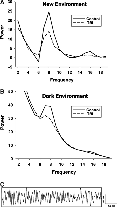
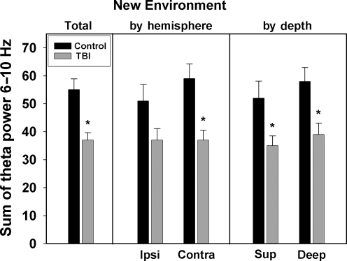
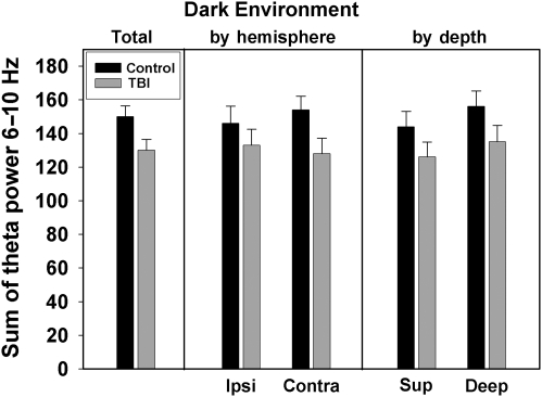
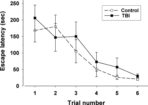
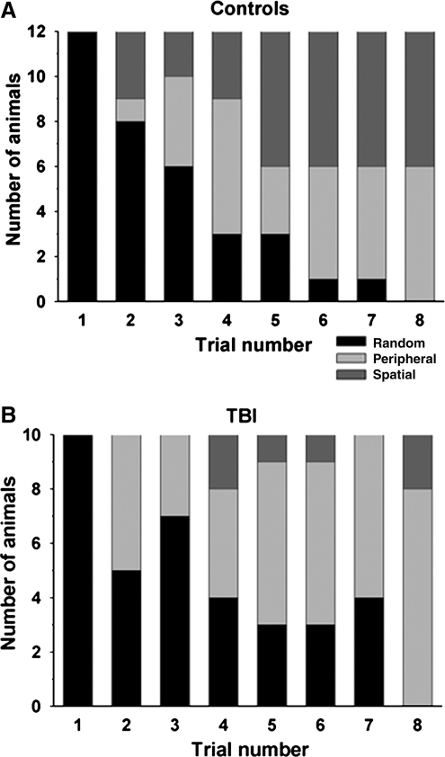
Chronic memory deficits are a major cause of morbidity following traumatic brain injury (TBI). In the rat, the hippocampal theta rhythm is a well-studied correlate of memory function. This study sought to investigate disturbances in hippocampal theta rhythm following lateral fluid percussion injury in the rat. A total of 13 control rats and 12 TBI rats were used. Electrodes were implanted in bilateral hippocampi and an electroencephalogram (EEG) was recorded while the rats explored a new environment, and also while navigating a modified version of the Barnes maze. Theta power and peak theta frequency were significantly attenuated in the injured animals. Further, injured rats were less likely to develop a spatial strategy for Barnes maze navigation compared to control rats. In conclusion, rats sustaining lateral fluid percussion injury demonstrated deficits in hippocampal theta activity. These deficits may contribute to the underlying memory problems seen in chronic TBI.
Figures
Similar articles
-
Septohippocampal Neuromodulation Improves Cognition after Traumatic Brain Injury.J Neurotrauma. 2015 Nov 15;32(22):1822-32. doi: 10.1089/neu.2014.3744. Epub 2015 Sep 2. J Neurotrauma. 2015. PMID: 26096267 Free PMC article.
-
Improved learning and memory with theta-burst stimulation of the fornix in rat model of traumatic brain injury.Hippocampus. 2014 Dec;24(12):1592-600. doi: 10.1002/hipo.22338. Epub 2014 Aug 21. Hippocampus. 2014. PMID: 25087862
-
Medial septal nucleus theta frequency deep brain stimulation improves spatial working memory after traumatic brain injury.J Neurotrauma. 2013 Jan 15;30(2):131-9. doi: 10.1089/neu.2012.2646. J Neurotrauma. 2013. PMID: 23016534
-
[Theta rhythm recorded in the hippocampal formation in vitro].Postepy Hig Med Dosw (Online). 2013 Jul 15;67:617-30. doi: 10.5604/17322693.1058537. Postepy Hig Med Dosw (Online). 2013. PMID: 24018425 Review. Polish.
-
Perspective: Hippocampal theta rhythm as a potential vestibuloacoustic biomarker of anxiety.Eur J Neurosci. 2025 Jan;61(1):e16641. doi: 10.1111/ejn.16641. Epub 2024 Dec 11. Eur J Neurosci. 2025. PMID: 39662900 Free PMC article. Review.
Cited by
-
Optogenetic Stimulation of CA1 Pyramidal Neurons at Theta Enhances Recognition Memory in Brain Injured Animals.J Neurotrauma. 2023 Nov;40(21-22):2442-2448. doi: 10.1089/neu.2023.0078. Epub 2023 Aug 23. J Neurotrauma. 2023. PMID: 37387400 Free PMC article.
-
A comparative study of human and rat hippocampal low-frequency oscillations during spatial navigation.Hippocampus. 2013 Aug;23(8):656-661. doi: 10.1002/hipo.22124. Epub 2013 Apr 29. Hippocampus. 2013. PMID: 23520039 Free PMC article.
-
Disrupted Hippocampal Theta-Gamma Coupling and Spike-Field Coherence Following Experimental Traumatic Brain Injury.bioRxiv [Preprint]. 2024 Sep 12:2024.05.30.596704. doi: 10.1101/2024.05.30.596704. bioRxiv. 2024. PMID: 39314320 Free PMC article. Preprint.
-
Affective, neurocognitive and psychosocial disorders associated with traumatic brain injury and post-traumatic epilepsy.Neurobiol Dis. 2019 Mar;123:27-41. doi: 10.1016/j.nbd.2018.07.018. Epub 2018 Jul 27. Neurobiol Dis. 2019. PMID: 30059725 Free PMC article. Review.
-
Frequency-Dependent Changes in Resting State Electroencephalogram Functional Networks after Traumatic Brain Injury in Piglets.J Neurotrauma. 2019 Sep 1;36(17):2558-2578. doi: 10.1089/neu.2017.5574. Epub 2019 May 23. J Neurotrauma. 2019. PMID: 30909806 Free PMC article.
References
-
- Bach M.E. Hawkins R.D. Osman M. Kandel E.R. Mayford M. Impairment of spatial but not contextual memory in CaMKII mutant mice with a selective loss of hippocampal LTP in the range of the theta frequency. Cell. 1995;81:905–915. - PubMed
-
- Barnes C. Memory deficits associated with senescence: a neurophysiological and behavioral study in the rat. J. Compar. Physiological Psychol. 1979;93:74–104. - PubMed
-
- Basar-Eroglu C. Demiralp T. Event-related theta oscillations: an integrative and comparative approach in the human and animal brain. Int. J. Psychophysiol. 2001;39:167–195. - PubMed
-
- Bland B.H. The physiology and pharmacology of hippocampal formation theta rhythms. Prog. Neurobiol. 1986;26:1–54. - PubMed
-
- Bramlett H.M. Green E.J. Dietrich W.D. Hippocampally dependent and independent chronic spatial navigational deficits following parasagittal fluid percussion brain injury in the rat. Brain Res. 1997;762:195–202. - PubMed
Publication types
MeSH terms
Grants and funding
LinkOut - more resources
Full Text Sources
