

Familial isolated clubfoot is associated with recurrent chromosome 17q23.1q23.2 microduplications containing TBX4
- PMID: 20598276
- PMCID: PMC2896772
- DOI: 10.1016/j.ajhg.2010.06.010
Familial isolated clubfoot is associated with recurrent chromosome 17q23.1q23.2 microduplications containing TBX4
Abstract
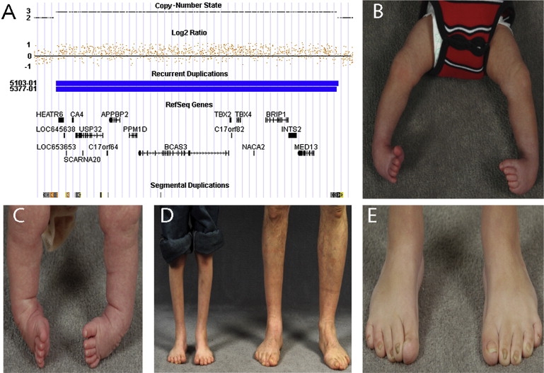
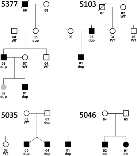
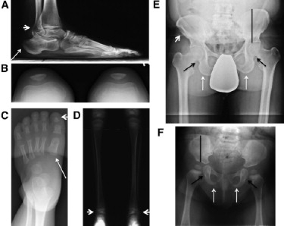
Clubfoot is a common musculoskeletal birth defect for which few causative genes have been identified. To identify the genes responsible for isolated clubfoot, we screened for genomic copy-number variants with the Affymetrix Genome-wide Human SNP Array 6.0. A recurrent chromosome 17q23.1q23.2 microduplication was identified in 3 of 66 probands with familial isolated clubfoot. The chromosome 17q23.1q23.2 microduplication segregated with autosomal-dominant clubfoot in all three families but with reduced penetrance. Mild short stature was common and one female had developmental hip dysplasia. Subtle skeletal abnormalities consisted of broad and shortened metatarsals and calcanei, small distal tibial epiphyses, and thickened ischia. Several skeletal features were opposite to those described in the reciprocal chromosome 17q23.1q23.2 microdeletion syndrome associated with developmental delay and cardiac and limb abnormalities. Of note, during our study, we also identified a microdeletion at the locus in a sibling pair with isolated clubfoot. The chromosome 17q23.1q23.2 region contains the T-box transcription factor TBX4, a likely target of the bicoid-related transcription factor PITX1 previously implicated in clubfoot etiology. Our result suggests that this chromosome 17q23.1q23.2 microduplication is a relatively common cause of familial isolated clubfoot and provides strong evidence linking clubfoot etiology to abnormal early limb development.
Copyright 2010 The American Society of Human Genetics. Published by Elsevier Inc. All rights reserved.
Figures
References
-
- Wynne-Davies R. Genetic and environmental factors in the etiology of talipes equinovarus. Clin. Orthop. Relat. Res. 1972;84:9–13. - PubMed
-
- Gurnett C.A., Boehm S., Connolly A., Reimschisel T., Dobbs M.B. Impact of congenital talipes equinovarus etiology on treatment outcomes. Dev. Med. Child Neurol. 2008;50:498–502. - PubMed
-
- Wynne-Davies R. Family studies and the cause of congenital club foot. Talipes equinovarus, talipes calcaneo-valgus and metatarsus varus. J. Bone Joint Surg. Br. 1964;46:445–463. - PubMed
-
- Lochmiller C., Johnston D., Scott A., Risman M., Hecht J.T. Genetic epidemiology study of idiopathic talipes equinovarus. Am. J. Med. Genet. 1998;79:90–96. - PubMed
Publication types
MeSH terms
Substances
Grants and funding
LinkOut - more resources
Full Text Sources
Molecular Biology Databases
