. 2010 Jul 9;87(1):129-38.
doi: 10.1016/j.ajhg.2010.06.002.

Mosaic uniparental disomies and aneuploidies as large structural variants of the human genome

Affiliations

Mosaic uniparental disomies and aneuploidies as large structural variants of the human genome

Benjamín Rodríguez-Santiago et al. Am J Hum Genet. .

Abstract

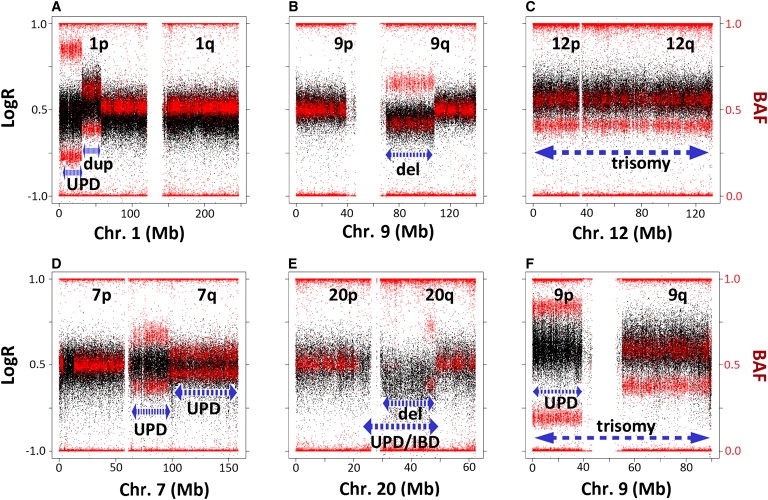
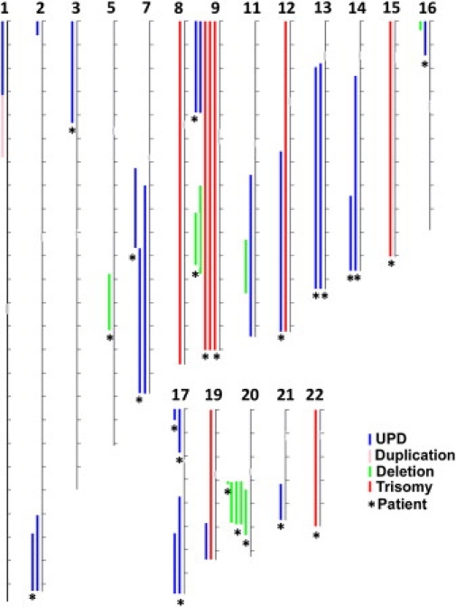
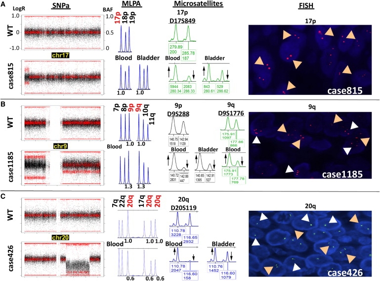
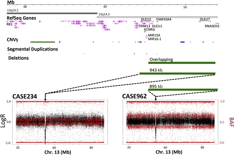
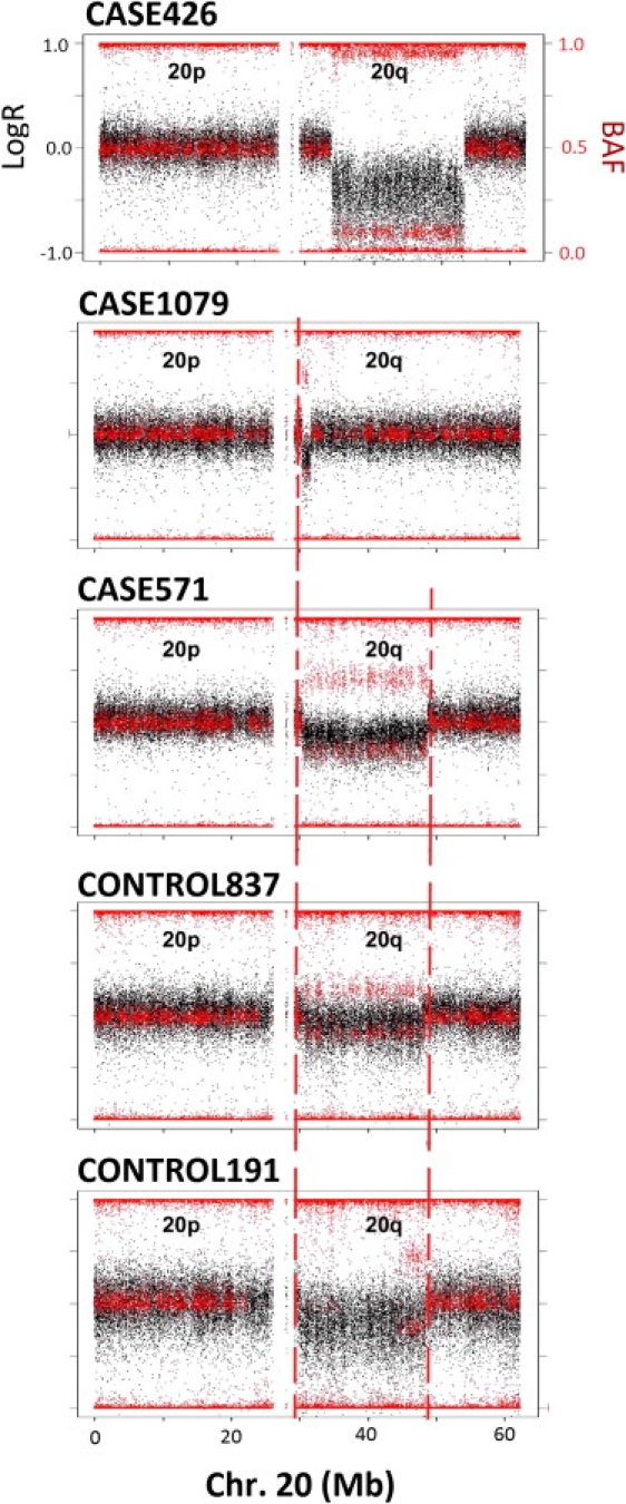
Mosaicism is defined as the coexistence of cells with different genetic composition within an individual, caused by postzygotic somatic mutation. Although somatic mosaicism for chromosomal abnormalities is a well-established cause of developmental and somatic disorders and has also been detected in different tissues, its frequency and extent in the adult normal population are still unknown. We provide here a genome-wide survey of mosaic genomic variation obtained by analyzing Illumina 1M SNP array data from blood or buccal DNA samples of 1991 adult individuals from the Spanish Bladder Cancer/EPICURO genome-wide association study. We found mosaic abnormalities in autosomes in 1.7% of samples, including 23 segmental uniparental disomies, 8 complete trisomies, and 11 large (1.5-37 Mb) copy-number variants. Alterations were observed across the different autosomes with recurrent events in chromosomes 9 and 20. No case-control differences were found in the frequency of events or the percentage of cells affected, thus indicating that most rearrangements found are not central to the development of bladder cancer. However, five out of six events tested were detected in both blood and bladder tissue from the same individual, indicating an early developmental origin. The high cellular frequency of the anomalies detected and their presence in normal adult individuals suggest that this type of mosaicism is a widespread phenomenon in the human genome. Somatic mosaicism should be considered in the expanding repertoire of inter- and intraindividual genetic variation, some of which may cause somatic human diseases but also contribute to modifying inherited disorders and/or late-onset multifactorial traits.


Figures

Figure 1
Figure 1
Examples of Different Types of Mosaic Rearrangements, Including Complex Rearrangements The plots show the signal intensity log R ratio (LogR) (black dots, scale on the left side) and B allele frequency (BAF) (red dots, scale on the right side) values along the entire chromosome carrying the rearrangements in selected samples. The length of the aberration is designated by the dashed blue lines; the type of rearrangement is annotated below. (A) Mosaic uniparental disomy (UPD) in distal 1p characterized by unchanged LogR and abnormal heterozygous BAF in the indicated interval; the interstitial mosaic duplication of the adjacent fragment shows elevated LogR (lower than the 0.2 cutoff for heterozygous duplication calling) and abnormal heterozygous BAF. (B) Large mosaic deletion in chromosome arm 9q showing decreased LogR and abnormal heterozygous BAF without complete loss of heterozygosity. (C) Mosaic trisomy 12 with a pattern similar to that of duplications along the entire chromosome. (D) Mosaic UPD with different degrees of mosaicism for two adjacent regions of chromosome arm 7q. (E) Large mosaic deletion in chromosome arm 20q (decreased LogR > −0.2 and abnormal heterozygous BAF). The complete loss of heterozygosity of a genomic region overlapping with the mosaic deletion suggests the presence of complete UPD or homozygosity resulting from identity by descent (IBD). (F) Mosaic trisomy for the entire chromosome 9 (increased LogR). The different B deviation value between 9p and 9q with identical LogR ratios in this sample also suggests the presence of UPD for 9p in addition to the trisomy.
Figure 2
Figure 2
Genomic Distribution of Mosaic Events Detected The illustration summarizes the chromosomal location and approximate size of all the mosaic events detected along the autosomal chromosomes: 23 UPDs (blue lines), 8 trisomies (red lines), one duplication (pink line), and 10 deletions (green lines). No alterations involving chromosomes 4, 6, 10, or 18 were found. Chromosomes are drawn to scale (tick marks indicate 10 Mb). Asterisks () indicate events found in a bladder cancer patient.
Figure 3
Figure 3
Representative Mosaic Rearrangements Validated by Independent Methods Validation was performed on the same source of DNA used for the SNP array (multiplex ligation-dependent probe amplification [MLPA] and microsatellites) as well as on DNA (microsatellites) and tumor tissue sections (fluorescence in situ hybridization [FISH]) from bladder tumor tissue. The plots at left show the LogR ratio (black dots) and BAF (red dots) values along an entire chromosome in selected samples (bottom) compared to the wild-type pattern (WT, top). (A) Segmental UPD of 17p terminal in case 815. MLPA (P060 panel) confirmed the disomic state at the UPD loci with no gain or loss of genetic material (1.0 relative peak height [RPH]), microsatellite analysis ratified the allelic imbalance both in blood and bladder tumor DNA reflected by an aberrant ratio between allelic peaks with respect to the control sample, and FISH showed two signals corresponding to disomy in all nuclei (tan arrowheads in panel at right). (B) Full trisomy 9 with additional UPD of 9p. MLPA confirmed a gain of genetic material at the trisomic locus (1.3 RPH), and microsatellite analysis ratified the allelic imbalance without detecting third alleles in both blood and bladder tumor DNA (shown by upward and downward arrows in middle panel). Right panel: FISH on bladder tumor tissue revealed a mosaic pattern of interphase cell nuclei with two (disomic, tan arrowheads), three (trisomic, white arrowheads; ∼30%), or four or more (tetrasomic or polysomic; ∼10%) signals. (C) Large (19.4 Mb) deletion-type copy-number variation (CNV) on chromosome 20q. MLPA confirmed a loss of genetic material at three loci within the interval, and microsatellite analysis ratified an allelic imbalance in both blood and bladder tumor DNA. Right panel: FISH on bladder tumor tissue revealed a mosaic pattern of interphase cell nuclei with either one (monosomic, white arrowheads) or two (disomic, tan arrowheads) signals. The percentage of cells carrying the rearrangement was higher in blood than in tumor cells in all three cases, as revealed by the greater degree of allelic imbalance.
Figure 4
Figure 4
Overlapping Rearrangements in Leukocyte DNA Samples from Two Patients with Bladder Cancer The plots on the bottom show the mosaic UPDs including almost the entire chromosome 13q found in blood DNA of two bladder cancer patients who also shared deletion-type heterozygous CNVs overlapping ∼833 kb at 13q14 (chr13 coordinates: 49539177–50372054). Analysis of the average LogR of the probes within the CNV yielded below-average LogR ratio for heterozygous deletion values in case 234 (left) and average values in case 962 (right); loss of heterozygosity was also found in case 234, whereas case 962 displayed values compatible with heterozygosity in 1/3 of SNPs within the interval, indicating that the disomic chromosome was the one carrying the CNV in case 234 whereas the other chromosome carried the CNV in case 962. The size of the deletions and overlapping fragment is represented by green bars illustrating their relative location with respect to the chromosome 13 ideogram and showing related genomic features: gene content (including distant RB1 gene), CNVs (Database of Genomic Variants, March 2010 version), and segmental duplications.
Figure 5
Figure 5
Overlapping Mitotic Interstitial Deletions of Chromosome 20q in Five Unrelated Individuals Aligned plots of LogR ratio (black) and BAF (red) values along the entire chromosome 20 of the five individuals, showing the rearrangements and the similar breakpoints (joined by dashed vertical red lines).

References

    1. Youssoufian H., Pyeritz R.E. Mechanisms and consequences of somatic mosaicism in humans. Nat. Rev. Genet. 2002;3:748–758. - PubMed
    1. Notini A.J., Craig J.M., White S.J. Copy number variation and mosaicism. Cytogenet. Genome Res. 2008;123:270–277. - PubMed
    1. Hsu L.Y., Kaffe S., Jenkins E.C., Alonso L., Benn P.A., David K., Hirschhorn K., Lieber E., Shanske A., Shapiro L.R. Proposed guidelines for diagnosis of chromosome mosaicism in amniocytes based on data derived from chromosome mosaicism and pseudomosaicism studies. Prenat. Diagn. 1992;12:555–573. - PubMed
    1. Hsu L.Y., Yu M.T., Richkind K.E., Van Dyke D.L., Crandall B.F., Saxe D.F., Khodr G.S., Mennuti M., Stetten G., Miller W.A., Priest J.H. Incidence and significance of chromosome mosaicism involving an autosomal structural abnormality diagnosed prenatally through amniocentesis: A collaborative study. Prenat. Diagn. 1996;16:1–28. - PubMed
    1. Rehen S.K., Yung Y.C., McCreight M.P., Kaushal D., Yang A.H., Almeida B.S., Kingsbury M.A., Cabral K.M., McConnell M.J., Anliker B. Constitutional aneuploidy in the normal human brain. J. Neurosci. 2005;25:2176–2180. - PMC - PubMed

Publication types

LinkOut - more resources