. 2010 Sep 30;405(2):289-99.
doi: 10.1016/j.virol.2010.05.019. Epub 2010 Jul 3.

Study of infectious virus production from HPV18/16 capsid chimeras

Affiliations

Study of infectious virus production from HPV18/16 capsid chimeras

Horng-Shen Chen et al. Virology. .

Abstract

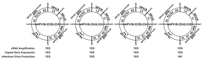
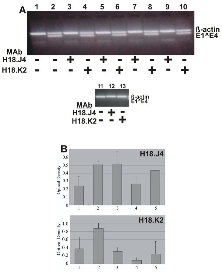
Using the HPV18 genome as the backbone, we exchanged the HPV18 L2 or L1 genes with those of HPV16. The intertypical exchange of HPV18 L1 with the HPV16 L1 produced genomes that efficiently replicated and produced infectious virus. Genomes containing an intertypical exchange of HPV18 L2 for the HPV16 L2 failed to produce infectious virus in multiple independently derived cell lines. Using chimeric constructs of individual capsid proteins, we identified a type-specific domain at the N-terminus of the HPV18L1 capsid protein, which interferes with its ability to cooperate with the HPV16 L2 protein to form infectious viral particles. Deletion of this domain allows for the cooperation of the HPV18 L1 protein and HPV16 L2 protein and production of infectious progeny. In addition, cooperation of this N-terminal HPV18 L1 deletion mutant protein with the wild-type HPV18 L2 protein efficiently replicates infectious virus but changes occur in the viral structure.


Figures

FIGURE 1
FIGURE 1
Late functions of viral mutants with nonoverlapping L2 and L1 ORFs. Late viral life cycle functions were analyzed for the four mutant HPV18 viruses containing nonoverlapping L2 and L1 ORFs; HPV18-L2(18)L1(18), HPV18-L2(16)L1(16), HPV18-L2(18)L1(16), and HPV18-L2(16)L1(18).
FIGURE 2
FIGURE 2
Western blot of HPV L1 capsid protein expression with the L1-specific MAb H18.E20. H18.E20 monoclonal antibody was used to detect L1 protein expression from chimeric HPV infected raft tissues. H18.E20 was raised against HPV18 L1 protein but cross-reacts with HPV16 L1 protein. Protein isolated from chimeric HPV18/CRPV virus infected raft tissues, expressing the CRPV L2 and L1 capsid proteins was used as a negative control. Protein isolated from wild-type HPV18 infected raft tissues was used as a positive control. Protein isolated from virus infected raft tissues were loaded, 90 or 120 μg to each well as labeled. Lanes 1–2: HPV18/CRPV infected tissues; Lanes 3–4: wild-type HPV18 infected tissues; Lanes 5–6: HPV18-L2(18)L1(18) infected tissues; Lanes 7–8: HPV18-L2(18)L1(16) infected tissues; Lanes 9–10: HPV18-L2(16)L1(18) infected tissues (cell line #1); Lanes 11–12: HPV18-L2(16)L1(18) infected tissues (cell line #2); and Lanes 13–14: HPV18-L2(16)L1(18) infected tissues (cell line #3). NOTE, that Lanes 9–14 represent three individually derived cell lines of the same mutant, HPV18-L2(16)L1(18). Molecular weight markers are on the left. The arrow indicates the position of the L1 protein.
FIGURE 3
FIGURE 3
Western blot of HPV L1 capsid protein expression with the L1-specific MAb H18.L9. H18.L9 monoclonal antibody was used to detect L1 protein expression from chimeric HPV infected raft tissues. H18.L9 was raised against HPV18 L1 protein but cross-reacts with HPV16 L1 protein. Protein isolated from chimeric HPV18/CRPV virus infected raft tissues, expressing the CRPV L2 and L1 capsid proteins was used as a negative control. Protein isolated from wild-type HPV18 infected raft tissues was used as a positive control. Protein isolated from virus infected raft tissues were loaded, 90 or 120 μg to each well as labeled. Lanes 1–2: HPV18/CRPV infected tissues; Lanes 3–4: wild-type HPV18 infected tissues; Lanes 5–6: HPV18-L2(18)L1(18) infected tissues; Lanes 7–8: HPV18-L2(18)L1(16) infected tissues; Lanes 9–10: HPV18-L2(16)L1(18) infected tissues (cell line #1); Lanes 11–12: HPV18-L2(16)L1(18) infected tissues (cell line #2); and Lanes 13–14: HPV18-L2(16)L1(18) infected tissues (cell line #3). NOTE, that Lanes 9–14 represent three individually derived cell lines of the same mutant, HPV18-L2(16)L1(18). Molecular weight markers are on the left. The arrow indicates the position of the L1 protein.
FIGURE 4
FIGURE 4
Western blot of HPV L2 capsid protein expression with the L2-specific MAb H16.4B4. H16.4B4 monoclonal antibody was used to detect L2 protein expression from chimeric HPV infected raft tissues. H16.4B4 was raised against HPV18 L2 protein but cross-reacts with HPV16 L2 protein. Protein isolated from chimeric HPV18/CRPV virus infected raft tissues, expressing the CRPV L2 and L1 capsid proteins was used as a negative control. Protein isolated from wild-type HPV18 infected raft tissues was used as a positive control. Protein isolated from virus infected raft tissues were loaded, 90 or 120 μg to each well as labeled. Lanes 1–2: HPV18/CRPV infected tissues; Lanes 3–4: wild-type HPV18 infected tissues; Lanes 5–6: HPV18-L2(18)L1(18) infected tissues; Lanes 7–8: HPV18-L2(18)L1(16) infected tissues; Lanes 9–10: HPV18-L2(16)L1(18) infected tissues (cell line #1); Lanes 11–12: HPV18-L2(16)L1(18) infected tissues (cell line #2); and Lanes 13–14: HPV18-L2(16)L1(18) infected tissues (cell line #3). NOTE, that Lanes 9–14 represent three individually derived cell lines of the same mutant, HPV18-L2(16)L1(18). Molecular weight markers are on the left. The arrow indicates the position of the L2 protein.
FIGURE 5
FIGURE 5
Capsid proteins L1 and L2 chimeric junctions. Shown are the approximate positions of the cloning junctions used for the L1 and L2 HPV18 and HPV16 chimeras. (A) Shown are the three-dimensional monomeric structure of L1 (left) and the amino acid sequences of HPV16 and HPV18 L1 proteins beginning at the consensus methionine (right) as described by Chen et al. (Chen et al., 2000). On the three-dimensional structure an oval shows the region on the F ß-sheet strand containing the chimeric junction. The position of the chimeric junction can also be seen of the two-dimensional sequence inside of the box. The positions of the ß-sheets (capital letter only for the three-dimensional structure and ß-capital letter for the sequence) and the helixes (h) are shown. (B) Shown are described functional regions of papillomavirus L2 proteins (Bossis et al., 2005; Fay et al., 2004; Florin et al., 2006; Florin et al., 2002; Gornemann et al., 2002; Heino, Zhou, and Lambert, 2000; Holmgren et al., 2005; Kamper et al., 2006; Kawana et al., 1999; Laniosz, Nguyen, and Meneses, 2007; Okun et al., 2001; Roden et al., 2001; Roden et al., 1994b; Yang et al., 2003a; Yang et al., 2003b; Zhou et al., 1994). The chimeric junction is shown at its position in the center of the L2 protein.
FIGURE 6
FIGURE 6
Southern (DNA) blot hybridization of chimeric HPV DNA-electroporated HFK cell lines grown in monolayer cultures. Cell lines were analyzed for the episomal maintenance, copy number, and integrity of the chimeric genomic DNA. Left Panels: The blots were probed with an HPV18-specific probe. Right Panels: The blots were stripped and reprobed with an HPV16-specific probe. Samples in lanes 1 and 4 were undigested, Samples in lanes 2 and 5 were digested with EcoRI, a single cutter of the chimeric genomes. Samples in lanes 3 and 6 were digested with BglII to separate the capsid genes (late ORFs) from the rest of the viral genome (early ORFs). Indicated are form I DNA (FI), form II DNA (FII), form II DNA (FIII), early ORFs (E), and late ORFs (L).
FIGURE 7
FIGURE 7
Infectivity assay of chimeric HPVs. Shown is a 2% agarose gel of nested RT-PCR-amplified HPV18 E1^E4 and ß-actin. All assays were done with use 1:20 dilution of the viral stock. Lane 1: negative control mock infected; Lane 2: positive control wildtype HPV18; Lane 3: HPV18-L2(16)L1(18/16); Lane 4: HPV18-L2(18)L1(18/16); Lane 5: HPV18-L2(18/16)L1(18); Lane 6: HPV18-L2(18)L1(16/18); Lane 7: HPV18-L2(16)L1(16/18); Lane 8: HPV18-L2(16/18)L1(18); Lane 9: HPV18-L2(16/18)L1(16); and Lane 10: HPV18-L2(18/16)L1(16). Positions of ß-actin and E1^E4 amplified sequences are shown on the right.
FIGURE 8
FIGURE 8
Papillomavirus L1 N-terminus amino acid alignment. Potential start sites for high-risk HPVs and the ‘consensus’ methionine are boxed.
FIGURE 9
FIGURE 9
(A) Neutralization of chimeric HPVs. Shown is a 2% agarose gel of nested RT-PCR-amplified HPV18 E1^E4 and ß-actin. Chimeric viruses were preincubated with the indicated MAb, H18.J4 or H18.K2. Lane 1: negative control, mock infected; Lane 2: positive control, wildtype HPV18; Lanes 3 and 4: HPV18-L2(18)L1(Δ18); Lanes 5 and 6: HPV18-L2(16)L1(Δ18); Lanes 7 and 8: HPV18-L2(18/16)L1(Δ18); Lanes 9 and 10: HPV18-L2(16/18)L1(Δ18); Lane 11: negative control, mock infected; Lanes 12 and 13: positive control, wildtype HPV18. Positions of ß-actin and E1^E4 amplified sequences are shown on the right. (B) Relative binding of conformation-dependent HPV18 L1-specific MAbs H18.J4 and H18.K2 to chimeric HPVs by ELISA assay. Relative binding for each chimeric HPV was defined as the difference between wells probed with (A) H18.J4 or (B) H18.K2 and wells probed with an irrelevant MAb. Values shown are means ± standard errors of the means. Lane 1: wildtype HPV18; Lane 2: HPV18-L2(18)L1(Δ18); Lane 3: HPV18-L2(16)L1(Δ18); Lane 4: HPV18-L2(18/16)L1(Δ18); and Lane 5: HPV18-L2(16/18)L1(Δ18).

Similar articles

Cited by

References

    1. Alam S, Conway MJ, Chen HS, Meyers C. The cigarette smoke carcinogen benzo[a]pyrene enhances human papillomavirus synthesis. J Virol. 2008;82(2):1053–8. - PMC - PubMed
    1. Becker KA, Florin L, Sapp C, Sapp M. Dissection of human papillomavirus type 33 L2 domains involved in nuclear domains (ND) 10 homing and reorganization. Virology. 2003;314(1):161–7. - PubMed
    1. Bedell MA, Hudson JB, Golub TR, Turyk ME, Hosken M, Wilbanks GD, Laimins LA. Amplification of human papillomavirus genomes in vitro is dependent on epithelial differentiation. J Virol. 1991;65(5):2254–60. - PMC - PubMed
    1. Bishop B, Dasgupta J, Klein M, Garcea R, Christensen N, Zhao R, Chen X. Crystal structures of four types of human papillomavirus L1 capsid proteins: understanding the specificity of neutralizing monoclonal antibodies. Journal of Biological Chemistry. 2007a;282(43):31803–31811. - PubMed
    1. Bishop B, Dasgupta J, Klein M, Garcea RL, Christensen ND, Zhao R, Chen XS. Crystal structures of four types of human papillomavirus L1 capsid proteins: understanding the specificity of neutralizing monoclonal antibodies. J Biol Chem. 2007b;282(43):31803–11. - PubMed

Publication types

MeSH terms

Substances