HB-EGF protects the lungs after intestinal ischemia/reperfusion injury
- PMID: 20599214
- PMCID: PMC2922487
- DOI: 10.1016/j.jss.2010.03.062
HB-EGF protects the lungs after intestinal ischemia/reperfusion injury
Abstract
Background: Acute respiratory distress syndrome continues to be a major source of morbidity and mortality in critically-ill patients. Heparin binding EGF-like growth factor (HB-EGF) is a biologically active protein that acts as an intestinal cytoprotective agent. We have previously demonstrated that HB-EGF protects the intestines from injury in several different animal models of intestinal injury. In the current study, we investigated the ability of HB-EGF to protect the lungs from remote organ injury after intestinal ischemia/reperfusion (I/R).
Methods: Mice were randomly assigned to one of the following groups: (1) sham-operated; (2) sham+HB-EGF (1200 microg/kg in 0.6 mL administered by intra-luminal injection at the jejuno-ileal junction immediately after identification of the superior mesenteric artery); (3) superior mesenteric artery occlusion for 45 min followed by reperfusion for 6 h (I/R); or (4) I/R+HB-EGF (1200 microg/kg in 0.6 mL) administered 15 min after vascular occlusion. The severity of acute lung injury was determined by histology, morphometric analysis and invasive pulmonary function testing. Animal survival was evaluated using Kaplan-Meier analysis.
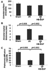
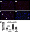
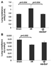
Results: Mice subjected to intestinal I/R injury showed histologic and functional evidence of acute lung injury and decreased survival compared with sham-operated animals. Compared with mice treated with HB-EGF (I/R+HB-EGF), the I/R group had more severe acute lung injury, and decreased survival.
Conclusion: Our results demonstrate that HB-EGF reduces the severity of acute lung injury after intestinal I/R in mice. These data demonstrate that HB-EGF may be a potential novel systemic anti-inflammatory agent for the prevention of the systemic inflammatory response syndrome (SIRS) after intestinal injury.
Copyright 2010 Elsevier Inc. All rights reserved.
Figures


Comment in
-
HB-EGF and mesenteric ischemia.J Surg Res. 2011 Jul;169(1):19-20. doi: 10.1016/j.jss.2010.09.045. Epub 2010 Oct 29. J Surg Res. 2011. PMID: 21316709 No abstract available.
Similar articles
-
Heparin-binding EGF-like growth factor decreases inflammatory cytokine expression after intestinal ischemia/reperfusion injury.J Surg Res. 2007 May 15;139(2):269-73. doi: 10.1016/j.jss.2006.10.047. Epub 2007 Feb 7. J Surg Res. 2007. PMID: 17291530 Free PMC article.
-
Heparin-binding epidermal growth factor-like growth factor gene disruption is associated with delayed intestinal restitution, impaired angiogenesis, and poor survival after intestinal ischemia in mice.J Pediatr Surg. 2008 Jun;43(6):1182-90. doi: 10.1016/j.jpedsurg.2008.02.053. J Pediatr Surg. 2008. PMID: 18558204 Free PMC article.
-
Heparin-binding epidermal growth factor-like growth factor attenuates acute lung injury and multiorgan dysfunction after scald burn.J Surg Res. 2013 Nov;185(1):329-37. doi: 10.1016/j.jss.2013.05.064. Epub 2013 Jun 12. J Surg Res. 2013. PMID: 23777985 Free PMC article.
-
Synergistic effects of HB-EGF and mesenchymal stem cells in a murine model of intestinal ischemia/reperfusion injury.J Pediatr Surg. 2013 Jun;48(6):1323-9. doi: 10.1016/j.jpedsurg.2013.03.032. J Pediatr Surg. 2013. PMID: 23845626 Free PMC article.
-
Heparin-binding epidermal growth factor-like growth factor and intestinal ischemia-reperfusion injury.Semin Pediatr Surg. 2004 Feb;13(1):2-10. doi: 10.1053/j.sempedsurg.2003.09.002. Semin Pediatr Surg. 2004. PMID: 14765365 Review.
Cited by
-
Germacrone ameliorates acute lung injury induced by intestinal ischemia-reperfusion by regulating macrophage M1 polarization and mitochondrial defects.Acta Biochim Biophys Sin (Shanghai). 2024 Oct 22;57(2):261-273. doi: 10.3724/abbs.2024164. Acta Biochim Biophys Sin (Shanghai). 2024. PMID: 39439416 Free PMC article.
-
Heparin-binding epidermal growth factor-like growth factor restores Wnt/β-catenin signaling in intestinal stem cells exposed to ischemia/reperfusion injury.Surgery. 2014 Jun;155(6):1069-80. doi: 10.1016/j.surg.2014.01.013. Epub 2014 Feb 6. Surgery. 2014. PMID: 24856127 Free PMC article.
-
Heparin-binding EGF-like growth factor (HB-EGF) protects the intestines from radiation therapy-induced intestinal injury.J Pediatr Surg. 2013 Jun;48(6):1316-22. doi: 10.1016/j.jpedsurg.2013.03.030. J Pediatr Surg. 2013. PMID: 23845625 Free PMC article.
-
Exploring transcriptomic mechanisms underlying pulmonary adaptation to diverse environments in Indian rams.Mol Biol Rep. 2024 Nov 1;51(1):1111. doi: 10.1007/s11033-024-10067-w. Mol Biol Rep. 2024. PMID: 39485559
-
Heparin-binding EGF-like growth factor (HB-EGF) therapy for intestinal injury: Application and future prospects.Pathophysiology. 2014 Feb;21(1):95-104. doi: 10.1016/j.pathophys.2013.11.008. Epub 2013 Dec 15. Pathophysiology. 2014. PMID: 24345808 Free PMC article.
References
-
- Rotstein OD. Pathogenesis of multiple organ dysfunction syndrome: gut origin, protection, and decontamination. Surg Infect. 2000;1:217–225. - PubMed
-
- Fink MP, Delude RL. Epithelial barrier dysfunction: a unifying theme to explain the pathogenesis of multiple organ dysfunction at the cellular level. Crit Care Clin. 2005;21:117–196. - PubMed
-
- Hotchkiss RS, Swanson PE, Freeman BD, Tinsley KW, Cobb JP, Matuschak GM, Buchman TG, Karl IE. Apoptotic cell death in patients with sepsis, shock, and multiple organ dysfunction. Crit Care Med. 1999;27:1230–1251. - PubMed
-
- Frutos-Vivar F, Ferguson ND, Esteban A. Epidemiology of acute lung injury and acute respiratory distress syndrome. Semin Respir Crit Care Med. 2006;27:327–336. - PubMed
MeSH terms
Substances
Grants and funding
LinkOut - more resources
Full Text Sources
Other Literature Sources

