Review
. 2010 Oct;21(10):628-36.
doi: 10.1016/j.tem.2010.06.002. Epub 2010 Jul 3.

Circadian clocks in the ovary

Affiliations
Review

Circadian clocks in the ovary

Michael T Sellix et al. Trends Endocrinol Metab. 2010 Oct.

Abstract

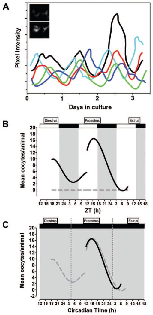
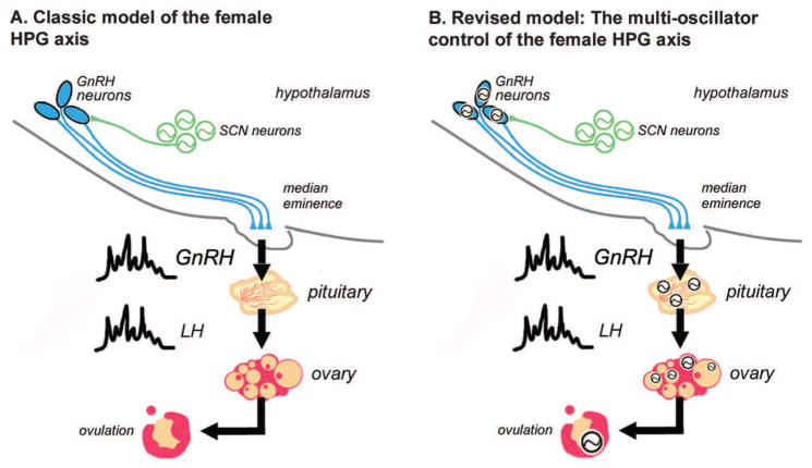
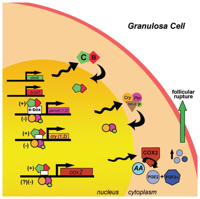
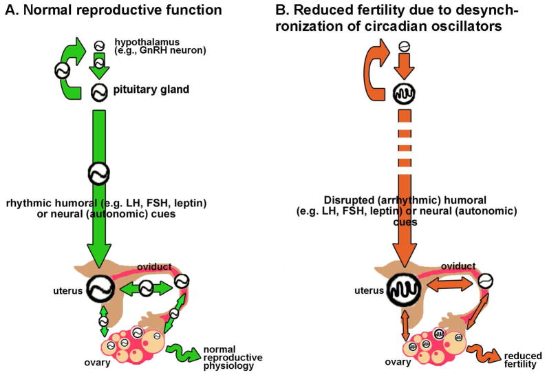
Clock gene expression has been observed in tissues of the hypothalamic-pituitary-gonadal (HPG) axis. Whereas the contribution of hypothalamic oscillators to the timing of reproductive biology is well known, the role of peripheral oscillators like those in the ovary is less clear. Circadian clocks in the ovary might play a role in the timing of ovulation. Disruption of the clock in ovarian cells or desynchrony between ovarian clocks and circadian oscillators elsewhere in the body may contribute to the onset and progression of various reproductive pathologies. In this paper, we review evidence for clock function in the ovary across a number of species and offer a novel perspective into the role of this clock in normal ovarian physiology and in diseases that negatively affect fertility.


Figures

Figure 1
Figure 1
A circadian clock in the rat ovary may contribute to the timing of ovulation. (a) Schematic representation showing circadian rhythms of period1-luciferase gene expression in individual granulosa/thecal cells. Inset graph: Images of trough (top panel) and peak (bottom panel) per1-luciferase gene expression in a representative granulosa/thecal cell recorded with an intensified CCD camera. Based on data from [7]. Schematic representation of (b) diurnal and (c) circadian rhythms of ovulation in response to exogenous LH in the absence of endogenous LH secretion. Animals housed under (b) a 12:12 L:D cycle or (c) constant dim light were injected with the GnRH receptor antagonist Cetrorelix on diestrus or proestrus to suppress endogenous LH secretion followed by timed injections of equine LH (solid black lines). LH-treatment during the subjective night on both diestrus and proestrus (L:D) or proestrus alone resulted in more frequent ovulation and significantly more oocytes/ovulation. Animals treated with sterile saline (gray dashed line in (b)) failed to ovulate regardless of injection time. The open and solid bars at the top of the figure indicate the light and dark portions of the L:D cycle. The solid gray background in (c) indicates that animals were maintained under constant dim light. Dashed gray lines in (f) are data from (b) re-plotted to emphasize the similarity of the results. Panels (b, c) modified from [60].
Figure 2
Figure 2
A paradigm shift: How the multi-oscillator HPG axis controls the timing of reproductive physiology. (a) Classic model for hypothalamic-pituitary-gonadal (HPG) axis regulation of mammalian reproductive physiology. On the afternoon of proestrus, the circadian clock in the SCN drives rhythmic release of GnRH that stimulates rhythmic secretion of LH. Circulating LH then stimulates the ovarian follicle leading to rupture and oocyte release. According to the classic model, the timing of events in this system is dependent solely on timing cues from the pacemaker neurons in the SCN. (b) A revised model for the “multi-oscillator circadian system” in the HPG axis emphasizing the existence of circadian oscillators in each component of the axis. Synchronization between SCN pacemakers, GnRH neurons, pituitary cells and ovarian cells is necessary for proper timing of physiological events controlled by the HPG. Based on this model, we hypothesize that disrupting circadian phase relationships among these tissues will have negative effects on reproductive physiology. Green circles containing sine waves represent SCN pacemaker neurons. Black circles containing sine waves represent rhythmic circadian clock gene expression within the cells of each tissue or region. Potential feedback relationships among the oscillators have been omitted for clarity. Gonadotrophin releasing hormone (GnRH), luteinizing hormone (LH), Suprachiasmatic nucleus (SCN).
Figure 3
Figure 3
The molecular clock in mammalian follicular cells may drive the expression of clock-controlled genes necessary for ovulation. Circadian clock genes, including activators [BMAL1 (B); CLOCK (C)] and repressors [period (per) and cryptochrome (cry)], are rhythmically expressed and phosphorylated by casein kinases in granulosa cells. Cyclooxygenase-2 (cox2), the rate limiting enzyme for prostanoid synthesis, has E-box sequences in its promoter region, and evidence suggests that CLOCK:BMAL1 heterodimers can bind to and activate cox2 transcription [78, 79]. Circadian rhythms of cox2 mRNA expression may result in rhythmic accumulation of COX2 enzyme. In turn, rhythms of COX2 enzyme expression may lead to rhythmic synthesis and accumulation of PGE2 and PGF2α. Increased levels of prostanoid synthesis, particularly in response to a surge in LH secretion, are associated with follicular rupture. Thus, circadian rhythms of cox2 mRNA synthesis might indirectly contribute to the timing of ovulation by establishing a ready pool of LH-inducible prostaglandins. Transactivation by BMAL1:CLOCK is indicated by (+); repression of BMAL1:CLOCK activity by PER:CRY is indicated by (−). Arrowheads attached to sine waves indicate rhythmic transcription/translation. Curved arrows indicate nuclear translocation. Abbreviations: arachidonic acid (AA); prostaglandin E2 (PGE2); prostaglandin F2α (PGF2α); phosphorylation (P); Casein kinase 1,2 (CK1,2).
Figure 4
Figure 4
A synchronized circadian system in the HPG may be necessary for rheostasis. (a) We suggest that rheostasis in the reproductive system depends on synchronization within and between circadian clocks in the HPG axis. For normal function circadian clocks in hypothalamic neuroendocrine cells (e.g. GnRH neurons), pituitary gland, uterus, oviduct and ovary (represented by circles containing a sine wave) must be appropriately synchronized. Regular temporal cues [indicated as green arrows in (a)] originating in the SCN and transduced by nervous and humoral outputs to the periphery to drive coordination of central and peripheral clocks. Temporal cues originating in peripheral oscillators (e.g. the uterus) may also alter timing in nearby peripheral oscillators. For clarity, several additional links and feedback circuits have been omitted from this schematic. (b) We hypothesize that disrupting normal synchronization, either by reducing the amplitude or robustness of circadian clocks in target tissues [indicated by circles containing abnormal waveforms] or by altering the rhythmicity or amplitude of temporal cues [indicated by red arrows] of central or peripheral origin may exacerbate (or even cause) diseases associated with reduced fertility.

Similar articles

Cited by

References

    1. Morse D, Cermakian N, Brancorsini S, Parvinen M, Sassone-Corsi P. No circadian rhythms in testis: Period1 expression is clock independent and developmentally regulated in the mouse. Mol Endocrinol. 2003;17:141. - PubMed
    1. Alvarez JD, Chen D, Storer E, Sehgal A. Non-cyclic and developmental stage-specific expression of circadian clock proteins during murine spermatogenesis. Biol Reprod. 2003;69:81–91. - PubMed
    1. Bebas P, Goodall CP, Majewska M, Neumann A, Giebultowicz JM, Chappell PE. Circadian clock and output genes are rhythmically expressed in extratesticular ducts and accessory organs of mice. Faseb J. 2009;23:523–533. - PMC - PubMed
    1. Karman BN, Tischkau SA. Circadian clock gene expression in the ovary: Effects of luteinizing hormone. Biol Reprod. 2006;75:624–632. - PubMed
    1. Fahrenkrug J, Georg B, Hannibal J, Hindersson P, Gras S. Diurnal rhythmicity of the clock genes Per1 and Per2 in the rat ovary. Endocrinology. 2006;147:3769–3776. - PubMed