

The molecular basis of corneal transparency
- PMID: 20599432
- PMCID: PMC3726544
- DOI: 10.1016/j.exer.2010.06.021
The molecular basis of corneal transparency
Abstract
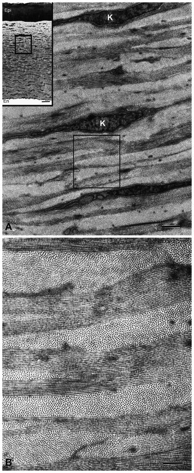
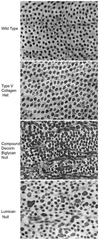
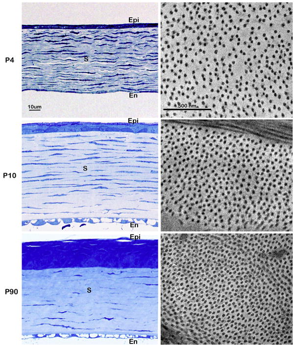
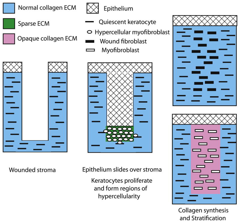
The cornea consists primarily of three layers: an outer layer containing an epithelium, a middle stromal layer consisting of a collagen-rich extracellular matrix (ECM) interspersed with keratocytes and an inner layer of endothelial cells. The stroma consists of dense, regularly packed collagen fibrils arranged as orthogonal layers or lamellae. The corneal stroma is unique in having a homogeneous distribution of small diameter 25-30 nm fibrils that are regularly packed within lamellae and this arrangement minimizes light scattering permitting transparency. The ECM of the corneal stroma consists primarily of collagen type I with lesser amounts of collagen type V and four proteoglycans: three with keratan sulfate chains; lumican, keratocan, osteoglycin and one with a chondroitin sulfate chain; decorin. It is the core proteins of these proteoglycans and collagen type V that regulate the growth of collagen fibrils. The overall size of the proteoglycans are small enough to fit in the spaces between the collagen fibrils and regulate their spacing. The stroma is formed during development by neural crest cells that migrate into the space between the corneal epithelium and corneal endothelium and become keratoblasts. The keratoblasts proliferate and synthesize high levels of hyaluronan to form an embryonic corneal stroma ECM. The keratoblasts differentiate into keratocytes which synthesize high levels of collagens and keratan sulfate proteoglycans that replace the hyaluronan/water-rich ECM with the densely packed collagen fibril-type ECM seen in transparent adult corneas. When an incisional wound through the epithelium into stroma occurs the keratocytes become hypercellular myofibroblasts. These can later become wound fibroblasts, which provides continued transparency or become myofibroblasts that produce a disorganized ECM resulting in corneal opacity. The growth factors IGF-I/II are likely responsible for the formation of the well organized ECM associated with transparency produced by keratocytes during development and by the wound fibroblast during repair. In contrast, TGF-beta would cause the formation of the myofibroblast that produces corneal scaring. Thus, the growth factor mediated synthesis of several different collagen types and the core proteins of several different leucine-rich type proteoglycans as well as posttranslational modifications of the collagens and the proteoglycans are required to produce collagen fibrils with the size and spacing needed for corneal stromal transparency.
Copyright (c) 2010 Elsevier Ltd. All rights reserved.
Figures

References
-
- Anseth A. Glycosaminoglycans in corneal regeneration. Exp Eye Res. 1961;1:122–127. - PubMed
-
- Beales MP, Funderburgh JL, Jester JV, Hassell JR. Proteoglycan synthesis by bovine keratocytes and corneal fibroblasts: maintenance of the keratocyte phenotype in culture. Invest Ophthalmol Vis Sci. 1999;40:1658–1663. - PubMed
-
- Beecher N, Carlson C, Allen BR, Kipchumba R, Conrad GW, Meek KM, Quantock AJ. An x-ray diffraction study of corneal structure in mimecan-deficient mice. Invest Ophthalmol Vis Sci. 2005;46:4046–4049. - PubMed
-
- Benedek GB. Theory of transparency of the eye. Appl Opt. 1971;10:459–473. - PubMed
-
- Bettelheim FA, Plessy B. The hydration of proteoglycans of bovine cornea. Biochim Biophys Acta. 1975;381:203–214. - PubMed
Publication types
MeSH terms
Substances
Grants and funding
LinkOut - more resources
Full Text Sources
Other Literature Sources
