. 2010 Jul 16;398(1):135-9.
doi: 10.1016/j.bbrc.2010.06.052. Epub 2010 Jun 17.

AMP-activated kinase (AMPK)-generated signals in malignant melanoma cell growth and survival

Affiliations

AMP-activated kinase (AMPK)-generated signals in malignant melanoma cell growth and survival

Jennifer Woodard et al. Biochem Biophys Res Commun. .

Abstract

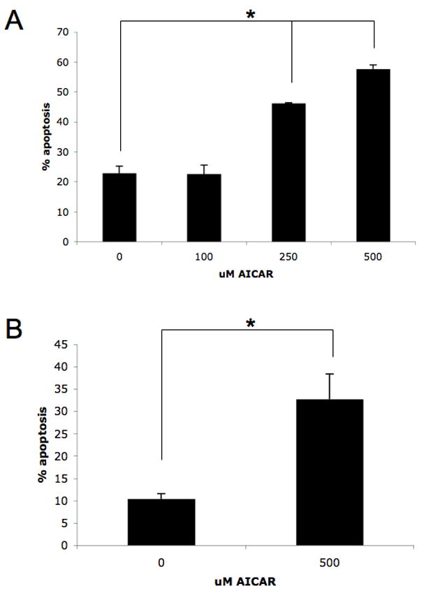
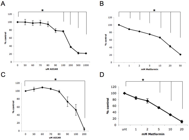
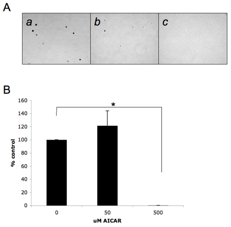
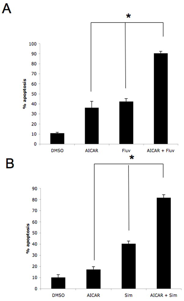
Extensive studies over the years have shown that the AMP-activated kinase (AMPK) exhibits negative regulatory effects on the activation of the mammalian target of rapamycin (mTOR) signaling cascade. We examined the potential involvement of AMPK in the regulation of growth and survival of malignant melanoma cells. In studies using the AMPK activators AICAR or metformin, we found potent inhibitory effects of AMPK activity on the growth of SK-MEL-2 and SK-MEL-28 malignant melanoma cells. Induction of AMPK activity was also associated with inhibition of the ability of melanoma cells to form colonies in an anchorage-independent manner in soft agar, suggesting an important role of the pathway in the control of malignant melanoma tumorigenesis. Furthermore, AICAR-treatment resulted in malignant melanoma cell death and such induction of apoptosis was further enhanced by concomitant statin-treatment. Taken together, our results provide evidence for potent inhibitory effects of AMPK on malignant melanoma cell growth and survival and raise the potential of AMPK manipulation as a novel future approach for the treatment of malignant melanoma.


Figures

Figure 1
Figure 1. Induction of pro-apoptotic effects by AMPK activators in malignant melanoma cells
A–B. SK-MEL-2 (A) or SK-MEL-28 (B) cells were incubated in the presence or absence of AICAR at the indicated concentrations for 96 hours. The induction of apoptosis was evaluated by flow cytometry using annexin V/propidium iodide staining. Data are expressed as percent apoptotic cells. Means + SE of three (A) or four (B) independent experiments for SK-MEL-2 or SK-MEL-28 cells, respectively, are shown. Paired t test analysis for the induction of apoptosis of SK-MEL-2 cells treated for 96 h with 250 μmol/L or 500 μmol/L versus control-treated cells showed two-tailed p values of 0.009 and 0.008, respectively. Paired t test analysis for the induction of apoptosis of SK-MEL-28 cells treated for 96 h versus control-treated cells showed a two-tailed p = 0.046.
Figure 2
Figure 2. Inhibitory effects of AMPK activators on malignant melanoma cell growth
A. SK-MEL-2 cells were treated for 4 days with solvent control or with the indicated concentrations of AICAR. Data are expressed as % control-treated cells and represent means + SE of 3 independent experiments. Paired t test analysis for the growth of SK-MEL-2 cells treated with 100 μmol/L AICAR, 200 μmol/L AICAR, 500 μmol/L AICAR, or 1000 μmol/L AICAR versus control-treated cells showed 2-tailed p values of 0.007, 0.0004, 0.001, and 0.0003, respectively. B. SK-MEL-2 cells were treated for 4 days with solvent control or with the indicated concentrations of metformin. Data are expressed as % control-treated cells and represent means + SE of 3 independent experiments. Paired t test analysis for the growth of SK-MEL-2 cells treated with 1 mmol/L metformin, 2 mmol/L metformin, 5 mmol/L metformin, 10 mmol/L metformin, 20 mmol/L metformin, or 50 mmol/L metformin versus control-treated cells showed 2-tailed p values of 0.012, 0.003, 0.001, 0.003, 0.002, and 0.0003, respectively. C. SK-MEL-28 cells were treated for 4 days with solvent control or with the indicated concentrations of AICAR. Data are expressed as % control-treated cells and represent means + SE of 3 independent experiments. Paired t test analysis for the growth of SK-MEL-28 cells treated with 200 μmol/L AICAR versus control-treated cells showed a 2-tailed p value = 0.002. D. SK-MEL-28 cells were treated for 4 days with solvent control or with the indicated concentrations of metformin. Data are expressed as % control-treated cells and represent means + SE of 3 independent experiments. Paired t test analysis for the growth of SK-MEL-28 cells treated with 5 mmol/L metformin, 10 mmol/L metformin, or 20 mmol/L metformin versus control-treated cells showed 2-tailed p values of 0.003, 0.0006, and 0.002, respectively.
Figure 3
Figure 3. Inhibitory effect of AICAR on melanoma cell anchorage-independent growth
A. Equal numbers of SK-MEL-28 cells were plated in a soft-agar assay system and treated with solvent control or AICAR (50 μmol/L or 500 μmol/L). Colony formation was analyzed after 14 days of culture. A representative area (20 x magnification) of each treatment point is shown (a–c). Cells were treated with solvent control (DMSO) (a), 50 μmol/L AICAR (b), or 500 μmol/L AICAR (c). Colonies were stained with crystal violet. B. Colonies were counted and results were expressed as % of solvent control-treated colonies. Data shown represent means + SE of 3 independent experiments. Paired t test analysis for the growth of SK-MEL-28 colonies treated with 500 μmol/L AICAR versus control-treated cells showed a 2-tailed p value = 2.56×10⁻⁷.
Figure 4
Figure 4. Enhanced pro-apoptotic responses in malignant melanoma cells by combinations of AICAR with statins
A. SK-MEL-28 cells were treated with solvent control, AICAR (500 μmol/L), fluvastatin (3 μmol/L), or the indicated combinations for ninety-six hours. Data are expressed as % apoptotic cells and represent means + SE of 3 independent experiments. Paired t test analysis for the induction of apoptosis of SK-MEL-28 cells treated with AICAR versus fluvastatin and AICAR showed a 2-tailed p value = 0.022. Paired t test analysis for the induction of apoptosis of SK-MEL-28 cells treated with fluvastatin versus fluvastatin and AICAR showed a 2-tailed p value = 0.006. B. SK-MEL-28 cells were treated with solvent control, AICAR (500 μmol/L), simvastatin (3 μmol/L), or the indicated combinations for ninety-six hours. Data are expressed as % apoptotic cells and represent means + SE of 3 independent experiments. Paired t test analysis for the induction of apoptosis of SK-MEL-28 cells treated with AICAR versus simvastatin and AICAR showed a 2-tailed p value = 0.006. Paired t test analysis for the induction of apoptosis of SK-MEL-28 cells treated with simvastatin versus simvastatin and AICAR showed a 2-tailed p value = 0.0007.

Similar articles

Cited by

References

    1. Hardie DG. The AMP-activated protein kinase pathway--new players upstream and downstream. J of Cell Sci. 2004;117(Pt 23):5479–5487. - PubMed
    1. Hardie DG, Hawley SA. AMP-activated protein kinase: the energy charge hypothesis revisited. Bioessays. 2001;23:1112–1129. - PubMed
    1. Hay N. The Akt-mTOR tango and its relevance to cancer. Cancer Cell. 2005;8:179–183. - PubMed
    1. Bhaskar PT, Hay N. The two TORCs and Akt. Dev Cell. 2007;12:487–502. - PubMed
    1. Gwinn DM, Shackelford DB, Egan DF, et al. AMPK phosphorylation of raptor mediates a metabolic checkpoint. Mol Cell. 2008;30:214–226. - PMC - PubMed

Publication types

MeSH terms