. 2010 Oct 15;80(8):1274-83.
doi: 10.1016/j.bcp.2010.06.043. Epub 2010 Jul 1.

Dexamethasone transcriptionally increases the expression of the pregnane X receptor and synergistically enhances pyrethroid esfenvalerate in the induction of cytochrome P450 3A23

Affiliations

Dexamethasone transcriptionally increases the expression of the pregnane X receptor and synergistically enhances pyrethroid esfenvalerate in the induction of cytochrome P450 3A23

Deshi Shi et al. Biochem Pharmacol. .

Abstract

The pregnane X receptor (PXR) is recognized as a key regulator for the induction of a large number of genes in drug metabolism and transport. The transactivation of PXR is enhanced by the glucocorticoid dexamethasone and the enhancement is linked to the induction of PXR in humans and rats. The present study was undertaken to determine the mechanism for the induction and ascertain the synergistic effect on the expression of CYP3A23, a rat PXR target. In primary hepatocytes, significant induction of PXR was detected as early as 2h after the treatment and the maximal induction occurred at 1 microM dexamethasone. Similar induction kinetics was observed in the hepatoma line H4-II-E-C3. The induction was abolished by actinomycin D and dexamethasone efficaciously stimulated the rat PXR promoter. In addition, dexamethasone synergized esfenvalerate (an insecticide and a PXR activator) in inducing CYP3A23 and stimulating the CYP3A23 promoter. The full promoter of CYP3A23 (-1445/+74) was activated in a similar pattern as the changes in PXR mRNA in response to dexamethasone, esfenvalerate and co-treatment. In contrast, different responding patterns were detected on the stimulation of the CYP3A23 proximal promoter. Synergistic stimulation was also observed on the CYP3A4-DP-Luc reporter, the human counterpart of CYP3A23. These findings establish that transactivation is responsible for the induction of rat PXR and the induction presents potential interactions with insecticides in a species-conserved manner. The different responding patterns among CYP3A23 reporters point to an involvement of multiple transcriptional events in the regulation of CYP3A23 expression by dexamethasone, esfenvalerate and both.


Figures

Fig. 1
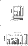
Fig. 1. Induction of rat PXR in primary hepatocytes by dexamethasone
Primary hepatocytes from Sprague-Dawley male rats (8-week old, n = 4) were isolated by a modified two-step collagenase digestion method. Hepatocytes were initially seeded and cultured in Williams’ E medium for 48 h with replacing the medium at 24 h. Thereafter, hepatocytes were cultured in dexamethasone-free medium for 16 h and then treated with dexamethasone at various concentrations (0–10 μM). Then hepatocytes were harvested for preparing cell lysates and total RNA. Total RNA was analyzed for the level of PXR mRNA by RT-qPCR and cell lysates (10 μg) were analyzed for the levels of PXR or CYP3A23 proteins by Western blotting. The RT-qPCR signals were normalized based on the abundance of Pol II mRNA. (A) Induction of PXR as a function of concentrations, and (B) Time-course study on the induction of PXR. The Ct value (threshold cycles) for DMSO controls in all time-points is around 25.65. *Statistically significant difference (p < 0.05).
Fig. 2
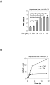
Fig. 2. Induction of rat PXR in H4-II-E-C3 cells by dexamethasone (A) Concentration-response study
H4-II-E-C3 cells were treated for 24 h with dexamethasone at various concentrations (0–10 μM), and total RNA was analyzed for the level of PXR mRNA by RT-PCR. Similarly the mRNA level was normalized based on the level of Pol II mRNA. Data were shown from three independent experiments. *Statistically significant difference from the vehicle control (p < 0.05). *Statistically significant difference (p < 0.05). (B) Time-course study H4-II-E-C3 cells were treated with dexamethasone at 50 nM and the cells were collected at 1, 2, 3, 4, 6 and 24 h. Similarly, total RNA was analyzed by qRT-PCR for the level of PXR or Tat mRNA by qRT-PCR, and the mRNA level was normalized based on the level of Pol II mRNA. The Ct value of PXR in DMSO control in all time-points is around 26.65.
Fig. 3
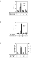
Fig. 3. Effect of actinomycin D, cycloheximide and RU486 on the dexamethasone-induced expression of PXR and Tat (A) Effect of actinomycin D
H4-II-E-C3 cells were seeded at a density of 5 × 10⁵ in 12-well plates. After an overnight incubation, the cells were treated with dexamethasone (50 nM), actinomycin D (Act D, 1 μM), or both for 6 h. The levels of PXR and Tat mRNA were determined by RT-qPCR. The signals were normalized according to the signal on Pol II mRNA. Letter “b” denotes statistical significance from letter “a”; letter “d” from letter “a” or letter “c”; and letter “a” from letter “c”. (B) Effect of RU486 H4-II-E-C3 cells were seeded as described above and subsequently treated with dexamethasone (50 nM), RU486 (0.1 μM), or both for 24 h. The levels of PXR and Tat mRNA were determined by RT-qPCR, and the signals were normalized according to the signal on Pol II mRNA. Data presented in this figure were assembled from three independent experiments. Letter “b” denotes statistical significance from letter “a”; and letter “d” from letter “c”. (C) Effect of cycloheximide on the dexamethasone-regulated expression of PXR and AGP H4-II-E-C3 cells were seeded as described above and subsequently treated with dexamethasone (50 nM), cycloheximide (CHX, 1 μM), or both for 24 h. The levels of PXR and Tat mRNA were determined by RT-qPCR, and the signals were normalized according to the signal on Pol II mRNA. Data presented in this figure were assembled from three independent experiments. Letter “b” denotes statistical significance from letter “a”; and letter “d” from letter “c”.
Fig. 4
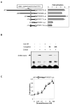
Fig. 4. Activation of the rat PXR promoter reporters by dexamethasone (A) Identification of dexamethasone response element in the PXR promoter
H4-II-E-C3 cells were seeded in 48-well plates at a density of 1.2 × 10⁵. After an overnight incubation, the cells were transiently transfected by GenJet with a mixture containing a reporter (50 ng) along with 5 ng of the tk-Renilla luciferase plasmid. After incubation at 37°C for 24 h (serum-free medium), the transfected cells were treated with dexamethasone (50 nM) or the same volume of DMSO for 48 h. Luciferase activities were determined with a Dual-Luciferase Reporter Assay System and the reporter activity was normalized based on the Renilla luminescence signal. The normalized reporter activities are expressed as fold of activation. (B) EMSA analysis Nuclear extracts (5 μg) from H4-II-E-C3 cells treated with dexamethasone (100 nM) were incubated with a biotinylated probe containing the putative GR element (0.1 pmol) for 20 min. In the competition assay, nuclear extracts were pre-incubated with the unlabeled probe at 5x or 20x excess for 5 min, and then incubated with the biotinylated probe. In disruption assay, nuclear extracts were incubated first with an antibody against glucocorticoid receptor-α on ice for 20 min and then with the biotinylated probe. The protein-DNA complexes were electrophoretically resolved and transferred to a Biodyne® nylon membrane. The biotinylated probe was located with streptavidin-conjugated horseradish peroxidase and chemiluminescent substrate. (C) Concentration-dependent activation of rPXR257-Luc H4-II-E-C3 cells were seeded and transfected as above. However, only the rPXR257-Luc was used in this study. The transfected cells were treated with dexamethasone at various concentrations (0–100 nM). Data presented in this figure were assembled from three independent experiments and each experiment was performed in triplicate. Bars with a different letter indicate statistically significant differences (p < 0.05).
Fig. 5
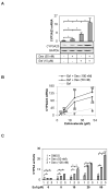
Fig. 5. Synergistic induction of CYP3A23 (A) Effect of dexamethasone on the induction of CYP3A23 by Esfenvalerate
Hepatocytes from Sprague-Dawley male rats (n = 4) were treated with dexamethasone (50 nM), esfenvalerate (10 μM), or both for 24 h. Total RNA was isolated and analyzed for the level of CYP3A23 mRNA by qRT-PCR. Cell lysates (10 μg) were analyzed for the level of CYP3A23 protein by Western blotting. *Statistically significant (p < 0.05). (B) Synergistic induction of CYP3A23 as a function of concentrations of dexamethasone and esfenvalerate Hepatocytes from Sprague-Dawley male rats (n = 4) were treated with various concentrations of dexamethasone (0–100 nM), esfenvalerate (0–50 μM), or both for 24 h. Total RNA was isolated and analyzed for the level of CYP3A23 mRNA (Left) or PXR (Right) by RT-qPCR. Bars with a letter “a” indicates statistically significant difference from DMSO-control; a letter “b” from the corresponding esfenvalerate treatment; and a letter “c” from esfenvalerate alone or esfenvalerate plus 50 nM dexamethasone (p < 0.05). (C) Enzymatic activity of CYP3A23 Hepatocytes from male rats (n = 4) were treated with various concentrations of dexamethasone (0–100 nM), esfenvalerate (0–50 μM) or both for 24 h. Hepatocytes were washed four times with phosphorous-buffed saline and cell lysates (20 μg) were then prepared. Lysates were analyzed for the oxidative activity with a P450-Glo CYP3A4 kit and the relative oxidation activity was determined with standard curves generated from various amounts of recombinant CYP3A4. *Statistically significant (p < 0.05), and bars with a different letter indicate statistically significant differences (p < 0.05) among data-points from DMSO- but different amounts of esfenvalerate. .
Fig. 6
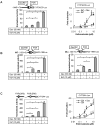
Fig. 6. Enhanced activation of CYP3A23 and CYP3A4 reporters
H4-II-E-C3 cells were seeded in 48-well plates at a density of 1.2 × 10⁵. After an overnight incubation, the cells were transiently transfected by GenJet with a mixture containing a reporter (50 ng) along with 5 ng of the tk-Renilla luciferase plasmid. After incubation at 37°C for 24 h (serum-free medium), the transfected cells were treated with dexamethasone (50 nM), esfenvalerate (10 μM), or both for 48 h. Alternatively, esfenvalerate was used at various concentrations (0–10 μM). Luciferase activities were determined with a Dual-Luciferase Reporter Assay System and the reporter activity was normalized based on the Renilla luminescence signal. (A) Activation of the CYP3A23-Luc reporter (basic + upstream regulatory sequence), (B) Activation of the CYP3A23-198Luc reporter (basic) or its mutant the CYP3A23-198mLuc reporter (basic with disrupted DexRE1), and (C) Activation of the CYP3A4-DP-Luc reporter. Data presented in this figure were assembled from three independent experiments and each experiment was performed in triplicate. *Statistical significance (Fig, 6B, Right of Figs. 6A and 6C) (p < 0.05) and bars with a different letter indicate statistically significant differences Figs. 6A and 6C (Right).

Similar articles

Cited by

References

    1. Ma X, Idle JR, Gonzalez FJ. The pregnane X receptor: from bench to bedside. Expert Opin Drug Metab Toxicol. 2008;4:895–908. - PMC - PubMed
    1. Zhou SF. Drugs behave as substrates, inhibitors and inducers of human cytochrome P450 3A4. Curr Drug Metab. 2008;9:310–322. - PubMed
    1. Goodwin B, Redinbo MR, Kliewer SA. Regulation of cyp3a gene transcription by the pregnane x receptor. Annu Rev Pharmacol Toxicol. 2002;42:1–23. - PubMed
    1. Watkins RE, Wisely GB, Moore LB, Collins JL, Lambert MH, Williams SP, Willson TM, Kliewer SA, Redinbo MR. The human nuclear xenobiotic receptor PXR: structural determinants of directed promiscuity. Science. 2001;292:2329–2333. - PubMed
    1. Watkins RE, Maglich JM, Moore LB, Wisely GB, Noble SM, Davis-Searles PR, Lambert MH, Kliewer SA, Redinbo MR. 2.1 A crystal structure of human PXR in complex with the St. John’s wort compound hyperforin. Biochemistry. 2003;42:1430–1438. - PubMed

Publication types

MeSH terms