. 2010 Oct 1;80(7):1021-32.
doi: 10.1016/j.bcp.2010.06.022. Epub 2010 Jun 23.

Cyclodextrin-complexed curcumin exhibits anti-inflammatory and antiproliferative activities superior to those of curcumin through higher cellular uptake

Affiliations

Cyclodextrin-complexed curcumin exhibits anti-inflammatory and antiproliferative activities superior to those of curcumin through higher cellular uptake

Vivek R Yadav et al. Biochem Pharmacol. .

Retraction in

Abstract

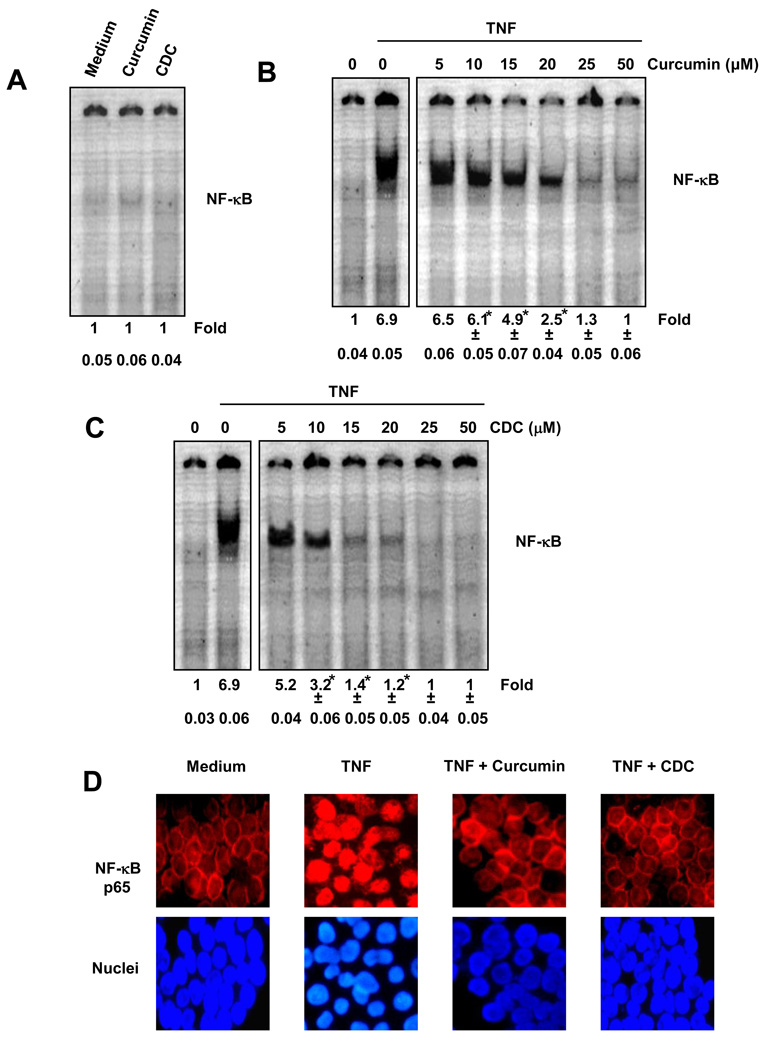
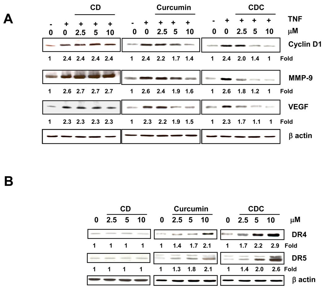
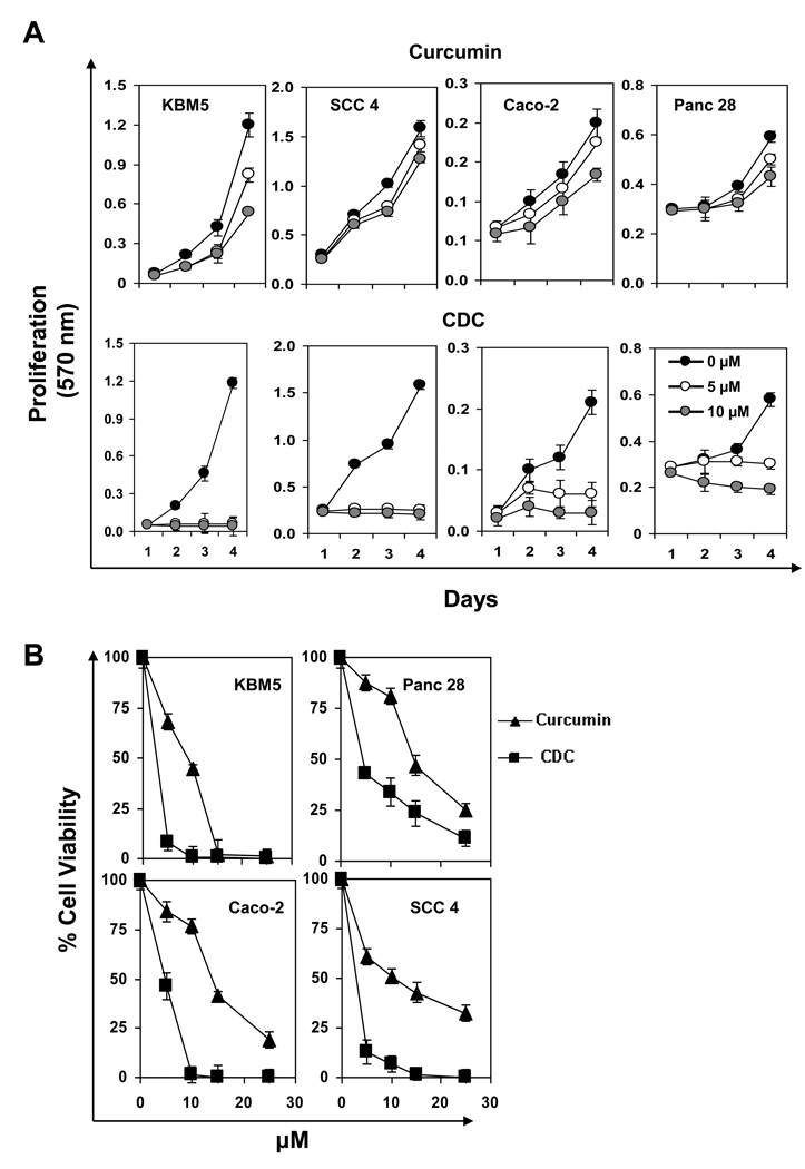
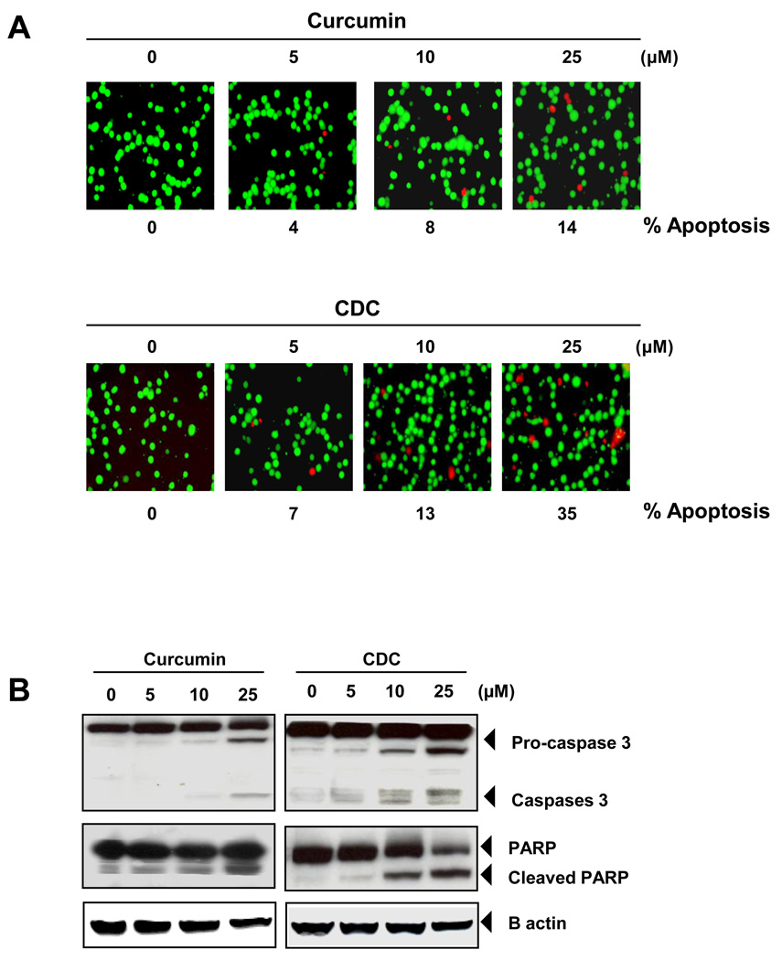
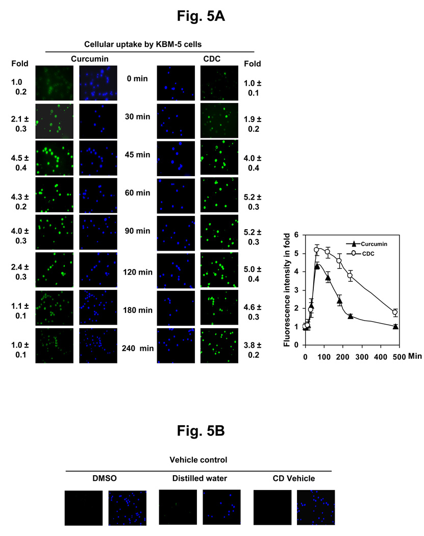
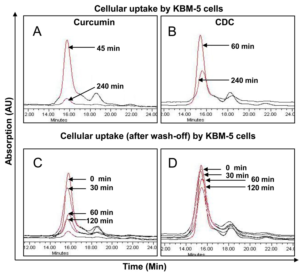
Curcumin, a yellow pigment present in the spice turmeric (Curcuma longa), has been linked with multiple beneficial activities, but its optimum potential is limited by poor bioavailability, in part due to the lack of solubility in aqueous solvents. To overcome the solubility problem, we have recently developed a novel cyclodextrin complex of curcumin (CDC) and examined here this compound for anti-inflammatory and antiproliferative effects. Using the electrophoretic mobility shift assay, we found that CDC was more active than free curcumin in inhibiting TNF-induced activation of the inflammatory transcription factor NF-kappaB and in suppressing gene products regulated by NF-kappaB, including those involved in cell proliferation (cyclin D1), invasion (MMP-9), and angiogenesis (VEGF). CDC was also more active than free curcumin in inducing the death receptors DR4 and DR5. Annexin V staining, cleavage of caspase-3 and PARP, and DNA fragmentation showed that CDC was more potent than free curcumin in inducing apoptosis of leukemic cells. Antiproliferative assays also demonstrated that CDC was more active than free curcumin in suppressing proliferation of various cancer cell lines. The cyclodextrin vehicle had no effect in these assays. Compared with free curcumin, CDC had a greater cellular uptake and longer half-life in the cells. Overall we demonstrated that CDC had superior attributes compared with free curcumin for cellular uptake and for antiproliferative and anti-inflammatory activities.


Figures

Figure 1
Figure 1
(A) CDC does not induce NF-κB activation in KBM-5 cells. KBM-5 cells (2 × 10⁶) were treated with the 50µM of curcumin or CDC for 4 h. Nuclear extracts were prepared and the NF-κB activity was examined by EMSA. (B and C) CDC is more potent than regular curcumin in inhibiting TNF-induced activation of NF-κB. KBM-5 cells (2 × 10⁶) were treated with the indicated concentrations of curcumin (B) or CDC (C) for 4 h. The cells were then incubated with 0.1 nM TNF for 30 min and analyzed for NF-κB activity by EMSA. The figures shown are representative of three independent experiments, and the numerical values expressed as mean ± SD are calculated from three independent experiments (D) Immunocytochemical analysis of TNF-induced p65 nuclear translocation. KBM-5 cells were incubated with 10 µM curcumin or 10 µM CDC for 4 h, treated with 0.1 nM TNF for 30 min, and then subjected to immunocytochemical analysis and examined in a fluorescence microscope. The figures shown are representative of three independent experiments. * indicate P < 0.05.
Figure 2
Figure 2
CDC is more potent than curcumin in inhibiting TNF-induced expression of NF-κB-regulated genes. KBM-5 cells (1 × 10⁶) were co-incubated with TNF (1 nM) and the indicated concentrations of curcumin or CDC for 24 h. The cells were harvested, and the expressions of cyclin D1, MMP-9, VEGF, and death receptors (DR4 and DR5) were analyzed by western blot. β-Actin was used as a loading control. The figures shown are representative of three independent experiments.
Figure 3
Figure 3
(A) Effect of CDC and curcumin on proliferation/survival as assessed by the 3-(4,5-dimethylthiazol-2-yl)-2,5-diphenyltetrazolium bromide (MTT) assay. CDC is more active than curcumin in suppressing proliferation at different days. (B) CDC showed more cytotoxic in different cancer cells. KBM-5 (human chronic myeloid leukemia), SCC-4 (human tongue squamous cancer), Caco-2 (human colonic carcinoma), and Panc-28 cells (pancreatic cancer) were incubated with different concentrations of either curcumin or CDC in for 72 h and then examined for cell viability by the MTT method. The results show the mean ± SD values.
Figure 4
Figure 4
CDC is more active than curcumin in inducing apoptosis. (A) KBM-5 cells (5000 cells/well) were incubated with curcumin or CDC at the indicated concentrations for 24 h. The cells were harvested and stained with Live/Dead assay reagent as per the manufacturer’s protocol as described in methods. The results shown are representative of three independent experiments. (B) CDC showed more apoptosis than curcumin. KBM-5 cells were treated with (5–25 µM) CD, curcumin or CDC for 24hr. Cleavage of caspase-3, and poly (ADP ribose) polymerase were determined by western blotting in whole-cell extracts of CD, Curcumin, and CDC treated cells. The results shown are representative of three independent experiments. (C) CDC showed more cell death than curcumin. KBM cells (2 × 10⁶/mL) were synchronized by incubation overnight in the absence of serum and then treated with (5–25 µM) CD, curcumin and CDC for 24hr, after which the cells were washed, fixed, stained with propidiumiodine and analyzed for DNA content by flow cytometry. (D) KBM-5 cells were treated with (5–25 µM) CD, curcumin and CDC for 24hr. Cell death was determined by fluorescence-activated cell sorting using annexin V/propidium iodide staining
Figure 4
Figure 4
CDC is more active than curcumin in inducing apoptosis. (A) KBM-5 cells (5000 cells/well) were incubated with curcumin or CDC at the indicated concentrations for 24 h. The cells were harvested and stained with Live/Dead assay reagent as per the manufacturer’s protocol as described in methods. The results shown are representative of three independent experiments. (B) CDC showed more apoptosis than curcumin. KBM-5 cells were treated with (5–25 µM) CD, curcumin or CDC for 24hr. Cleavage of caspase-3, and poly (ADP ribose) polymerase were determined by western blotting in whole-cell extracts of CD, Curcumin, and CDC treated cells. The results shown are representative of three independent experiments. (C) CDC showed more cell death than curcumin. KBM cells (2 × 10⁶/mL) were synchronized by incubation overnight in the absence of serum and then treated with (5–25 µM) CD, curcumin and CDC for 24hr, after which the cells were washed, fixed, stained with propidiumiodine and analyzed for DNA content by flow cytometry. (D) KBM-5 cells were treated with (5–25 µM) CD, curcumin and CDC for 24hr. Cell death was determined by fluorescence-activated cell sorting using annexin V/propidium iodide staining
Figure 5
Figure 5
(A) Cellular uptake of curcumin and CDC. KBM-5 cells (1 × 10⁶) were incubated with curcumin or CDC at concentrations of 10 µM. The cells were harvested at the indicated times, and the cellular uptake was determined by fluorescence microscopy as described in methods and blue color Hoechst’s staining showed cell viability. The results shown are representative of three independent experiments. (B) The control KBM-5 cells (2 × 10⁶) treated with DMSO and water. The results shown are representative of three independent experiments.
Figure 6
Figure 6
Cellular release of CDC is slower than curcumin. KBM-5 cells (1 × 10⁶) were incubated with curcumin or CDC at concentrations of 10 µM for 45 and 60 min, washed two times with PBS, and again incubated at 37°C. The cells were harvested at the indicated times, and the cellular uptake was determined by fluorescence microscopy as described in methods. The results shown are representative of three independent experiments.
Figure 7
Figure 7
Overlapped chromatograms of curcumin levels in the cells after treatment with curcumin or CDC. Chromatograms A and B show cellular uptake of curcumin and CDC, respectively, at different time points during treatment with 10µM concentration. Chromatograms C and D show curcumin levels in the cells at different time intervals after wash of the cells. The cell density was 5×10⁶/mL of KBM-5 cells.

Similar articles

Cited by

References

    1. Ravindran J, Prasad S, Aggarwal BB. Curcumin and cancer cells: how many ways can curry kill tumor cells selectively? Aaps J. 2009;11:495–510. - PMC - PubMed
    1. Tharakan ST, Inamoto T, Sung B, Aggarwal BB, Kamat AM. Curcumin potentiates the antitumor effects of gemcitabine in an orthotopic model of human bladder cancer through suppression of proliferative and angiogenic biomarkers. Biochem Pharmacol. 2009 - PMC - PubMed
    1. Kunnumakkara AB, Anand P, Aggarwal BB. Curcumin inhibits proliferation, invasion, angiogenesis and metastasis of different cancers through interaction with multiple cell signaling proteins. Cancer Lett. 2008;269:199–225. - PubMed
    1. Ralhan R, Pandey MK, Aggarwal BB. Nuclear factor-kappa B links carcinogenic and chemopreventive agents. Front Biosci (Schol Ed) 2009;1:45–60. - PubMed
    1. Aggarwal S, Ichikawa H, Takada Y, Sandur SK, Shishodia S, Aggarwal BB. Curcumin (diferuloylmethane) down-regulates expression of cell proliferation and antiapoptotic and metastatic gene products through suppression of IkappaBalpha kinase and Akt activation. Mol Pharmacol. 2006;69:195–206. - PubMed

Publication types