. 2010 Sep 15;345(2):117-32.
doi: 10.1016/j.ydbio.2010.06.028. Epub 2010 Jun 28.

Nonmuscle myosin II is required for cell proliferation, cell sheet adhesion and wing hair morphology during wing morphogenesis

Affiliations

Nonmuscle myosin II is required for cell proliferation, cell sheet adhesion and wing hair morphology during wing morphogenesis

Josef D Franke et al. Dev Biol. .

Abstract

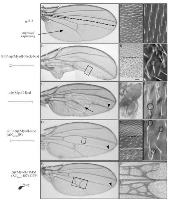
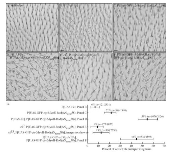
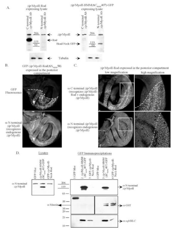
Metazoan development involves a myriad of dynamic cellular processes that require cytoskeletal function. Nonmuscle myosin II plays essential roles in embryonic development; however, knowledge of its role in post-embryonic development, even in model organisms such as Drosophila melanogaster, is only recently being revealed. In this study, truncation alleles were generated and enable the conditional perturbation, in a graded fashion, of nonmuscle myosin II function. During wing development they demonstrate novel roles for nonmuscle myosin II, including in adhesion between the dorsal and ventral wing epithelial sheets; in the formation of a single actin-based wing hair from the distal vertex of each cell; in forming unbranched wing hairs; and in the correct positioning of veins and crossveins. Many of these phenotypes overlap with those observed when clonal mosaic analysis was performed in the wing using loss of function alleles. Additional requirements for nonmuscle myosin II are in the correct formation of other actin-based cellular protrusions (microchaetae and macrochaetae). We confirm and extend genetic interaction studies to show that nonmuscle myosin II and an unconventional myosin, encoded by crinkled (ck/MyoVIIA), act antagonistically in multiple processes necessary for wing development. Lastly, we demonstrate that truncation alleles can perturb nonmuscle myosin II function via two distinct mechanisms--by titrating light chains away from endogenous heavy chains or by recruiting endogenous heavy chains into intracellular aggregates. By allowing myosin II function to be perturbed in a controlled manner, these novel tools enable the elucidation of post-embryonic roles for nonmuscle myosin II during targeted stages of fly development.


Figures

Figure 1
Figure 1. Schematics of the zip/MyoII constructs encoded by truncation alleles
In bold is the name of each construct, and the protein it encodes, used throughout this paper. In parenthesis is the amino acids of zip/MyoII that each includes. The predicted molecular weight is also given. To the right of each construct name is the base pair (bp) and amino acid (AA) sequence including the region of zip/MyoII (in bold), GFP (italics, if present) and the linker between these two proteins (plain). A rough schematic of the quaternary structure that each fragment is expected to assemble under high salt conditions is given. In the schematic, the zip/MyoII heavy chains are diagrammed as white or black globular and coiled structures. Both associated light chains are indicated by arrows (ELC is head proximal and RLC is head distal). For GFP fusion proteins, GFP is diagrammed as a barrel structure. The boxed region in lower right describes two new polyclonal antisera generated against zip/MyoII (N-terminal and C-terminal). The amino acid residues for each antigen and the antisera designation are given. The relative location of each antigen, with respect to the entire heavy chain, is indicated in the GFP-zip/MyoII (top) schematic.
Figure 2
Figure 2. Expression of zip/MyoII truncation alleles identifies novel roles for zip/MyoII in wing development
(A,A’,A”) Images of a control (w1118) wing. (A) The dashed line indicates the approximate boundary between the anterior (above dotted line) and posterior (below dotted line; engrailed expressing) compartments of the wing. (A’ and A”) High magnification SEM images of a wing showing single wing hairs protruding from the apical side of each cell and pointing uniformly toward the tip of the wing. (B,B’,B”) Expression of GFP-zip/MyoII-Neck-Rod. (B) Image of a whole mount wing showing a typical example of a loss of tissue in the posterior compartment (the extent of wing tissue lost varies). (B’) A high magnification brightfield image showing both the wild type (above dotted line) and GFP-zip/MyoII-Neck-Rod expressing regions (below dotted line) showing a multiple wing hair and split hair phenotypes. This region is approximated by the boxed region in B. (B”) High magnification SEM image showing a multiple wing hair phenotype and some branched hairs (circles). (C,C’,C”) Expression of zip/MyoII-Rod. (C) Image of a whole mount zip/MyoII-Rod-expressing wing containing a wing blister (arrow). (C’) A low magnification, brightfield image of the wing blister phenotype in a newly eclosed fly. (C”) High magnification SEM image showing both a multiple wing hair phenotype and a branched hair phenotype (circle) – both phenotypes were observed in posterior compartment cells regardless of their location relative to the blister. (D,D’,D”) Expression of GFP-zip/MyoII-Rod(ΔNterm58). (D) Image of a whole mount wing. (D’) High magnification SEM image showing the multiple wing hair phenotype (the dotted line indicates the anterior/posterior compartment boundary). (D”) A higher magnification SEM image shows branched and multiple wing hair phenotypes (circles). (E,E’,E”) Increased expression of zip/MyoII-HMM(ΔCterm407)-GFP caused crossvein phenotypes. (E) Image of a whole mount wing with ectopic crossveins associated with the posterior cross vein (see boxed region). Additional crossvein tissue was sometimes associated with the anterior crossvein and the distal tip of the longitudinal, L4 vein is often perturbed (arrowhead). (E’ and E”) High magnification brightfield images of ectopic and expanded crossveins.
Figure 3
Figure 3. Several types of actin-based cellular protrusions exhibit similar phenotypes when zip/MyoII function is perturbed
(A–C) Expression of GFP-zip/MyoII-Neck-Rod in the dorsal compartment of the wing disc by apterous-GAL4, in an otherwise wild-type background, caused thoracic defects. (A) SEM image shows three examples of two microchaetae (bristles) extending from a single socket (arrow). Setae, small hairs protruding from epidermal cells on the thorax also exhibit multiple cellular protrusions and branched phenotypes (circle). (B) SEM image showing different types of microchaetae (three wild-type microchaetae (arrows), multiple microchaetae and branched microchaetae). (C) SEM image of a single microchaetae splitting approximately two-thirds of the way to the bristle tip (arrowhead). (D–F) Expression of GFP-zip/MyoII-Rod(ΔNterm58) with apterous-GAL4, in an otherwise wild type background, causes similar defects. (D) Brightfield image showing multiple cellular protrusions occurs for both setae and microchaetae. (E and F) SEM images showing branching occurs in microchaetae (arrow in E) and setae (arrow in F). Scale bars in all panels are 25μm.
Figure 4
Figure 4. zip/MyoII function is required in the dorsal wing epithelial sheet for adhesion
SEM images of flies specifically expressing GFP-zip/MyoII-Neck-Rod in the dorsal epithelial sheet of the wing. (A,C,E) Wings appear as balloon-like appendages on either side of the fly (C and E are different views of the same wing). The two epithelial sheets only contact one another along the wing margin (arrows in A, B and C). (B) A higher magnification image of the boxed region in A shows the wing margin (arrow) where the two sheets meet. (D) A high magnification image of the boxed region in C shows that the dorsal, GFP-zip/MyoII-Neck-Rod expressing cells exhibit a multiple wing hair phenotype and a complete loss of vein and crossvein patterning (see panels A and C for lower magnification images). (E) Image of the ventral side of a wing. The ventral sheet retains a significant amount of wild-type wing vein patterning as both veins and crossveins (arrowheads) are present. (F) A high magnification image of the boxed region in E shows that wing hairs from ventral cells also have wild type morphology. The scale bars in A, C and E are 100μm and the scale bars in B, D, F are 25μm.
Figure 5
Figure 5. Expression of GFP-zip/MyoII-Rod(ΔNterm58) reveals a genetic interaction between zip/MyoII and Frizzled and between zip/MyoII and ck/MyoVIIA
(A) Brightfield image of individual wing hairs pointing in the same direction on a wild-type (w1118) wing. (B) Overexpression of Frizzled (w; P[en-GAL4]/+; P[UAS-Fz]/+) caused a mild multiple wing hair phenotype (for all mutant genotypes quantification is shown in G). (C) Expression of GFP-zip/MyoII-Rod(ΔNterm58) caused a multiple wing hair phenotype (w; P[en-GAL4]/+, P[UAS-GFP-zip/MyoII-Rod(ΔNterm58)/+). (D) Expression of both Frizzled and GFP-zip/MyoII-Rod(ΔNterm58) caused enhancement of the multiple wing hair phenotype and perturbed polarity (w; P[en-GAL4],P[UAS-GFP-zip/MyoII-Rod(ΔNterm58)/+; P[UAS-Fz]/+). (E) Expression of GFP-zip/MyoII-Rod(ΔNterm58) in a ck7 (a severe ck/MyoVIIA allele, Kiehart et al., 2004) heterozygous background suppresses the GFP-zip/MyoII-Rod(ΔNterm58) induced a multiple wing hair phenotype (w; P[en-GAL4], P[UAS-GFP-zip/MyoII-Rod(ΔNterm58)/ck7). Similar suppression was observed in a different, severe ck/MyoVIIA background (ck13, see G). (F) Expression of GFP-zip/MyoII-Rod(ΔNterm58) along with a functional UAS-GFP-ck/MyoVIIA transgene (Todi et al., 2005), resulted in the enhancement of the multiple wing hair phenotype (w; P[en-GAL4], P[UAS-GFP-zip/MyoII-Rod(ΔNterm58)/+; P[UAS-GFP-ck/MyoVIIA]/+). (G) Quantification of the percent multiple wing hair for the different genotypes. For each genotype indicated (y-axis) the percent multiple wing hair (x-axis) is provided. Among wings of the same genotype, some variability in the percent of multiple wing hairs was observed. A horizontal bar indicates the range of the multiple wing hair phenotype observed for wings of each genotype. Scale bar is 10μm.
Figure 6
Figure 6. Domain requirements for proper zip/MyoII temporal and spatial localization
Each zip/MyoII GFP-fusion protein (see schematic to the left of each time series) was ubiquitously expressed by sqh-Gal4 in an otherwise wild-type background unless specified. (A-C, video 1) GFP-zip/MyoII localized to the leading edge of dorsal-most cells (arrow) in a bars on a string distribution along both lateral epidermis sheets. The inset in A shows a high magnification image, from a later point in this time series, of a single cell with a large GFP-zip/MyoII aggregate next to the nucleus. (D-F, video 2) GFP-zip/MyoII-Neck-Rod similarly localized to the leading edge of dorsal-most cells. (G-I, video 3) To determine if GFP-zip/MyoII-Neck-Rod localization was dependent on endogenous zip/MyoII, we examined the distribution of GFP-zip/MyoII-Neck-Rod in zip/MyoII-deficient embryos (w; P[UAS-GFP-zip/MyoII-Neck-Rod], zip2 / P[sqh-GAL4], sp zip1). Here, GFP-zip/MyoII-Neck-Rod was unable to localize and persist at the leading edge of dorsal-most cells. Toward the end of closure, a small tear (arrow in I) in the embryo occurs that ultimately results in a dorsal hole. (H-L, video 4) The GFP-zip/MyoII-Rod(ΔNterm58) protein exhibited faint localization to the leading edge of dorsal-most cells. In instances where it was observed at the leading edge it failed to form bars within cells and failed to persist throughout the course of closure. (M-O, video 5) zip/MyoII-HMM(ΔCterm407)-GFP failed to exhibit any identifiable subcellular localization in any cell examined. Instead, this construct remained diffuse in the cytoplasm. Insets in B, E, H, K and N show a high magnification view of the boxed region in their respective panels. The scale bar in A is 20μm and applies to all large panels. The scale bar in the inset in B is 5μm and applies to all insets.
Figure 7
Figure 7. Antibody studies provide evidence for the mechanism by which myosin truncation alleles perturb myosin function
(A) Immunoblots demonstrate that specific N-terminal (AA 1-704) and C-terminal (AA 1631-1972) zip/MyoII antisera were generated. Larval lysates containing endogenous, zip/MyoII and either zip/MyoII-Rod (left) or zip/MyoII-HMM(ΔCterm407)-GFP (right) were probed with both antisera. By immunoblot, the C-terminal zip/MyoII antisera recognized zip/MyoII-Rod and endogenous zip/MyoII but not zip/MyoII-HMM(ΔCterm407)-GFP and the N-terminal zip/MyoII antisera recognized zip/MyoII-HMM(ΔCterm407)-GFP and endogenous zip/MyoII, but not zip/MyoII-Rod. Tubulin was probed as a loading control. (B) Expression of GFP-zip/MyoII-Rod(ΔNterm58) resulted in large aggregates within cells (top panel in B). The distribution of endogenous zip/MyoII (N-terminal antisera staining; bottom panel in B) was altered in just the GFP-zip/MyoII-Rod(ΔNterm58) expressing cells – a large amount of endogenous myosin was localized to the GFP-zip/MyoII-Rod(ΔNterm58) aggregates. In the control, anterior compartment cortical staining for zip/MyoII is observed. (C) Similar results were observed in disc cells expressing the zip/MyoII-Rod construct. The high magnification panels show the boxed region in the panel to their left. Scale bars are 10μm. (D) Both zip/MyoII-HMM(ΔCterm407)-GFP and GFP-zip/MyoII-Neck-Rod bind light chains. Larval lysates expressing zip/MyoII-HMM(ΔCterm407)-GFP or GFP-zip/MyoII-Neck-Rod were prepared under high salt conditions, to eliminate associations with endogenous zip/MyoII. As a control, lysates were prepared from larvae expressing GFP-Moesin. (Lysates blot) Each lysate contained a comparable amount of endogenous zip/MyoII and verified the presence of zip/MyoII-HMM(ΔCterm407)-GFP in the appropriate lysate. (GFP Immunopreciptiations lanes 1-3) Fusion proteins were immunoprecipitated using GFP antibodies and the immunoprecipitants were probed for zip/MyoII, moesin and sqh/RLC. The α-zip/MyoII and α-moesin blots verify that the immunoprecipitations were specific for their respective antigens and that no detectable endogenous zip/MyoII was pulled down. sqh/RLC was present in both the zip/MyoII-HMM(ΔCterm407)-GFP and GFP-zip/MyoII-Neck-Rod immunoprecipitations, but not the GFP-moe immunoprecipitation. (GFP Immunoprecipitations, lanes 4-6) To determine if both light chains (sqh/RLC and mlc-c/ELC) bound both constructs, similar larval lysates were prepared and bacterially purified GST-sqh/RLC and GST-mlc-c/ELC were added to each lysate. Similar immunoprecipitations were performed and each was probed for zip/MyoII, GST and sqh/RLC. The doublet in the GST blot shows that both GST-sqh/RLC and GST-mlc-c/ELC bound each zip/MyoII fragment. As a control, neither GST-fusion was found in the GFP-moe immunoprecipitation. Endogenous sqh/RLC was again observed in both the zip/MyoII-HMM(ΔCterm407)-GFP and GFP-zip/MyoII-Neck-Rod immunoprecipitations.

Similar articles

Cited by

References

    1. Adler PN. Planar signaling and morphogenesis in Drosophila. Dev Cell. 2002;2:525–35. - PubMed
    1. Adler PN, et al. Tissue polarity points from cells that have higher Frizzled levels towards cells that have lower Frizzled levels. Curr Biol. 1997;7:940–9. - PubMed
    1. Barros CS, et al. Drosophila nonmuscle myosin II promotes the asymmetric segregation of cell fate determinants by cortical exclusion rather than active transport. Dev Cell. 2003;5:829–40. - PubMed
    1. Bertet C, et al. Myosin-dependent junction remodelling controls planar cell intercalation and axis elongation. Nature. 2004;429:667–71. - PubMed
    1. Blair SS. Wing vein patterning in Drosophila and the analysis of intercellular signaling. Annu Rev Cell Dev Biol. 2007;23:293–319. - PubMed

Publication types

LinkOut - more resources