. 2010 Aug 15;344(2):1011-25.
doi: 10.1016/j.ydbio.2010.06.022. Epub 2010 Jun 23.

Genome-wide analysis of germ cell proliferation in C.elegans identifies VRK-1 as a key regulator of CEP-1/p53

Affiliations

Genome-wide analysis of germ cell proliferation in C.elegans identifies VRK-1 as a key regulator of CEP-1/p53

Katherine Waters et al. Dev Biol. .

Abstract

Proliferating germ cells in Caenorhabditiselegans provide a useful model system for deciphering fundamental mechanisms underlying the balance between proliferation and differentiation. Using gene expression profiling, we identified approximately 200 genes upregulated in the proliferating germ cells of C. elegans. Functional characterization using RNA-mediated interference demonstrated that over forty of these factors are required for normal germline proliferation and development. Detailed analysis of two of these factors defined an important regulatory relationship controlling germ cell proliferation. We established that the kinase VRK-1 is required for normal germ cell proliferation, and that it acts in part to regulate CEP-1(p53) activity. Loss of cep-1 significantly rescued the proliferation defects of vrk-1 mutants. We suggest that VRK-1 prevents CEP-1 from triggering an inappropriate cell cycle arrest, thereby promoting germ cell proliferation. This finding reveals a previously unsuspected mechanism for negative regulation of p53 activity in germ cells to control proliferation.


Figures

Fig. 1
Fig. 1
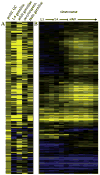
Identification of genes expressed in proliferating germ cells. (A) A clustergram of the 202 genes with expression >1.8×, p<.01 in glp-1(gf) animals relative to wild type (left column, prolif. GC), using microarray data from Reinke et al. (2004) to further characterize the expression profiles of these genes in germline development (all columns to the right). Each column header describes the stage and/or tissue in which expression of that gene is expected. Yellow indicates higher expression, blue indicates lower expression. For the oocyte/sperm comparison, yellow indicates high expression in oocytes, blue is high expression in sperm. (B) The developmental expression in whole animals of the same 202 genes. Data also from Reinke et al. (2004).
Fig. 2
Fig. 2
Functional categories of genes expressed in proliferating germ cells. Pie chart showing the predicted functions of the 202 genes. See sheet two of Supplemental File 1 for assignment of functional categories to individual genes.
Fig. 3
Fig. 3
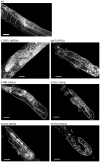
Example germline RNAi phenotypes for proliferating germ cell factors. Whole animal DAPI staining showing one gonad arm from a wild type control and six animals undergoing RNAi for genes from different functional categories (nucleolar: C15H11.9, E02H1.1, K12H4.3; unknown: C50B8.3, Y47G6A.9; and chaperone: daf-21). Visible region of gonad outlined in white; asterisk indicates distal tip. Scale bars represent 20 μm.
Fig. 4
Fig. 4
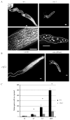
Loss of vrk-1 disrupts germline development. (A) Gonads dissected from wild type and vrk-1 mutant adult animals were stained with DAPI. Chromatin morphology is magnified in bottom panel to show condensed chromatin in vrk-1 mutant germ cells. Scale bars represent 20 μm. (B) Gonads dissected from rrf-1 mutants undergoing either control (L4440) or vrk-1 RNAi, and stained with DAPI. Scale bars same as in A. (C) The average number of germ cells per gonad arm was quantified in developing hermaphrodites from wild type and vrk-1 mutants. Error bars represent standard deviation. Student’s t test p values for L3, L4, and adult data are <2.0×10⁻⁵ (asterisks).
Fig. 5
Fig. 5
Apoptosis does not account for the vrk-1 mutant germ cell defects. (A) SYTO-12 staining was quantified in wild type and vrk-1 mutant germ cells. Error bars represent standard error values. Student’s t test p<2.0×10⁻⁵ (asterisks). (B) Dissected gonads from ced-4, vrk-1, and ced-4;vrk-1 mutants were stained with DAPI. Gonads outlined in white. Scale bars represent 20 μm.
Fig. 6
Fig. 6
Proliferation is reduced in the germ line of vrk-1 mutants. (A) The number of nuclei in mitosis was determined by counting pH3-positive nuclei in dissected gonads from wild type and vrk-1 mutants (n>20). Error bars represent standard error values. Student’s t test p values are <0.005 (asterisks). (B) BrdU-positive E. coli were fed to hermaphrodites at progressively later stages of development (blue lines). Green lines indicate stage at which BrdU was first fed and successfully incorporated in the vrk-1 mutant, while red indicates the stages at which feeding commenced but BrdU was no longer incorporated. (C) Gonads from adult wild type (left column) and vrk-1 mutant (right column) hermaphrodites exposed to BrdU-positive bacteria at each developmental stage were dissected and stained with antibodies against BrdU (red) and DAPI (DNA). Total number of gonads analyzed, and percentage with BrdU incorporation, is listed in the bottom right corner of each panel. Scale bars represent 20 μm.
Fig. 6
Fig. 6
Proliferation is reduced in the germ line of vrk-1 mutants. (A) The number of nuclei in mitosis was determined by counting pH3-positive nuclei in dissected gonads from wild type and vrk-1 mutants (n>20). Error bars represent standard error values. Student’s t test p values are <0.005 (asterisks). (B) BrdU-positive E. coli were fed to hermaphrodites at progressively later stages of development (blue lines). Green lines indicate stage at which BrdU was first fed and successfully incorporated in the vrk-1 mutant, while red indicates the stages at which feeding commenced but BrdU was no longer incorporated. (C) Gonads from adult wild type (left column) and vrk-1 mutant (right column) hermaphrodites exposed to BrdU-positive bacteria at each developmental stage were dissected and stained with antibodies against BrdU (red) and DAPI (DNA). Total number of gonads analyzed, and percentage with BrdU incorporation, is listed in the bottom right corner of each panel. Scale bars represent 20 μm.
Fig. 7
Fig. 7
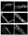
vrk-1 mutant germ nuclei have defects in both mitosis and interphase. A transgenic strain expressing both GFP:PCNA (green) and H2B:mCherry (pink) to mark chromatin was crossed to vrk-1 mutants. Live animals were mounted for examination of GFP:PCNA localization at L3, L4, and adult. Gonads are outlined in white, and the distal tip is indicated by an asterisk. The far right panels are insets from the adult stage, indicated by the dotted square. In the vrk-1 mutant germ line, examples of enlarged interphase cells are indicated by arrows, whereas a nucleus with more condensed DNA is marked by an arrowhead, in the inset. Scale bars represent 20 μm.
Fig. 8
Fig. 8
VRK-1 is expressed in the nuclei of almost all germ cells. (A) Gonads dissected from adult wild type animals were stained with DAPI (blue) to visualize DNA and antibodies against VRK-1 (red). The gonad is outlined in white. Spermatids lacking VRK-1 staining are indicated with an arrow, and the distal tip is marked by an asterisk. The scale bar represents 20 μm. (B) High power images of the distal region of adult gonads (white outline) expressing functional GFP:VRK-1 (green) and H2B:mCherry (red). Arrow shows discrete puncta of H2B:mCherry and GFP:VRK-1 colocalization. The arrowhead points to a ring of GFP:VRK-1 surrounding the DNA, presumably the nuclear envelope. Scale bars represent 5 μm.
Fig. 9
Fig. 9
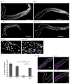
Loss of cep-1 rescues germline proliferation defects of vrk-1 mutant hermaphrodites. (A) DAPI-stained gonads from adult wild type, cep-1, vrk-1, and cep-1;vrk-1 hermaphrodites. Scale bars represent 50 μm. (B) High power view showing chromatin morphology. Scale bars represent 5 μm. (C) The number of pH3-positive nuclei in adults was quantified for each genotype (n>20/genotype). Student’s t test p values between cep-1 and cep-1;vrk-1 backgrounds are not significant (NS); p value between vrk-1 and cep-1;vrk-1 backgrounds is <1.8×10⁻⁹. (D) Wild type, cep-1, vrk-1 and cep-1;vrk-1 mutants were exposed to BrdU at the L4 stage and stained for BrdU incorporation (red) as adults. DNA was stained with DAPI (blue). Scale bars represent 10 μm.
Fig. 10
Fig. 10
A trans-heterozygous cep-1 mutant partially rescues the vrk-1 mutant phenotype. DAPI staining of dissected gonads from hermaphrodite adults of the genotype indicated in each panel. Gonads are outlined in white and regions within dashed boxes are magnified in the right column. (A) cep-1(lg12501) did not improve the germline size or DNA morphology of vrk-1 mutants. (B) A trans-heterozygous cep-1(lg12501)/cep1(gk138) background displayed partial rescue of vrk-1 germline defects. (C) A single copy of cep-1 (gk138) in a heterozygous background failed to rescue vrk-1 mutant defects. Scale bars represent 20 μm.
Fig. 11
Fig. 11
The DNA damage checkpoint is not activated in vrk-1 mutants. Whole animals were fixed and stained with DAPI to visualize DNA. The vrk-1 mutant germ line phenotype is not improved by the loss of each of the required members of the DNA damage checkpoint pathway: mrt-2, clk-2, or hus-2. The cep-1; vrk-1 mutant is shown as a control for rescue of the vrk-1 mutant phenotype. Gonads are outlined in white; asterisks indicate the distal tip in each image.

Similar articles

Cited by

References

    1. Aggarwal BD, Calvi BR. Chromatin regulates origin activity in Drosophila follicle cells. Nature. 2004;430:372–376. - PubMed
    1. Aladjem MI, Spike BT, Rodewald LW, Hope TJ, Klemm M, Jaenisch R, Wahl GM. ES cells do not activate p53-dependent stress responses and undergo p53-independent apoptosis in response to DNA damage. Curr Biol. 1998;8:145–155. - PubMed
    1. Berry LW, Westlund B, Schedl T. Germ-line tumor formation caused by activation of glp-1, a Caenorhabditis elegans member of the Notch family of receptors. Development. 1997;124:925–936. - PubMed
    1. Boehme KA, Blattner C. Regulation of p53–insights into a complex process. Crit Rev Biochem Mol Biol. 2009;44:367–392. - PubMed
    1. Bosl GJ, Motzer RJ. Testicular germ-cell cancer. N Engl J Med. 1997;337:242–253. - PubMed

Publication types

Substances