

Differential susceptibility to experimental glaucoma among 3 mouse strains using bead and viscoelastic injection
- PMID: 20599961
- PMCID: PMC2954410
- DOI: 10.1016/j.exer.2010.06.018
Differential susceptibility to experimental glaucoma among 3 mouse strains using bead and viscoelastic injection
Abstract
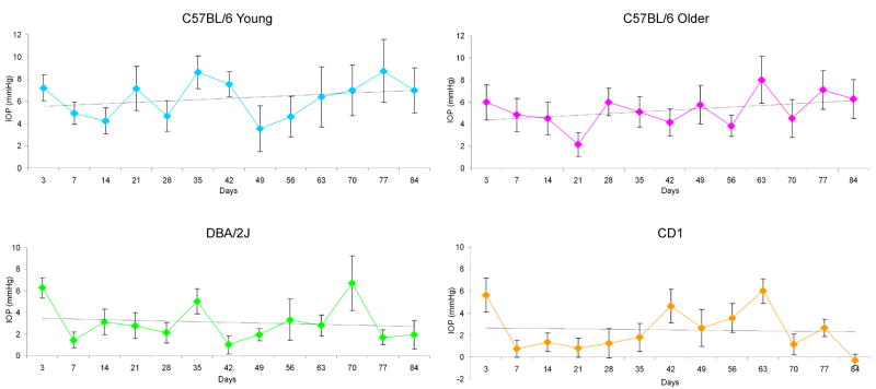
The purpose of this experiment was to test the susceptibility to retinal ganglion cell (RGC) axon loss and RGC layer cell loss from experimental glaucoma among 3 mouse strains, and between younger and older mice. We obstructed the mouse aqueous outflow channels by injecting 2 microL of 6 mum diameter, polystyrene beads followed by 3 microL of viscoelastic solution into the anterior chamber with a glass micropipette. We evaluated intraocular pressure (IOP) and damage to RGC as measured by optic nerve axon counts and RGC layer neuron counts in 3 strains of young mice (2 month old C57BL/6, DBA/2J, and CD1) and 10 month C57BL/6 mice. Bead and viscoelastic injection produced IOP elevation at >or=1 time point in 94.1% of eyes (112/119), with mean IOP difference from fellow eyes of 4.4 +/- 3.0 mmHg. By 6-12 weeks, injected eyes were 10.8% longer and 7.6% wider (p < 0.0001). Young DBA/2J and C57BL/6 eyes increased axial length significantly more than young CD1 or older C57BL/6 (all p <or= 0.02). RGC layer and axon loss was greatest in CD1 mice, significantly more than the other groups (p from 0.04 to <0.0001). Young C57BL/6 eyes elongated more and lost more RGC layer cells than older C57BL/6 mice (p = 0.02 and 0.01, respectively). With this mouse glaucoma model, there was differential susceptibility to ocular elongation and RGC layer and axon damage among mouse strains and by age. Factors that determine sensitivity to RGC injury can be studied using transgenic mouse strains with inducible models.
Copyright (c) 2010 Elsevier Ltd. All rights reserved.
Figures

Similar articles
-
The effects of anesthesia, mouse strain and age on intraocular pressure and an improved murine model of experimental glaucoma.Exp Eye Res. 2012 Jun;99(1):27-35. doi: 10.1016/j.exer.2012.04.006. Epub 2012 Apr 25. Exp Eye Res. 2012. PMID: 22554836 Free PMC article.
-
Mice with an induced mutation in collagen 8A2 develop larger eyes and are resistant to retinal ganglion cell damage in an experimental glaucoma model.Mol Vis. 2012;18:1093-106. Epub 2012 May 1. Mol Vis. 2012. PMID: 22701298 Free PMC article.
-
Lack of neuroprotection against experimental glaucoma in c-Jun N-terminal kinase 3 knockout mice.Exp Eye Res. 2011 Apr;92(4):299-305. doi: 10.1016/j.exer.2011.01.006. Epub 2011 Jan 25. Exp Eye Res. 2011. PMID: 21272576 Free PMC article.
-
Assessment of retinal ganglion cell damage in glaucomatous optic neuropathy: Axon transport, injury and soma loss.Exp Eye Res. 2015 Dec;141:111-24. doi: 10.1016/j.exer.2015.06.006. Epub 2015 Jun 9. Exp Eye Res. 2015. PMID: 26070986 Review.
-
Mouse models of retinal ganglion cell death and glaucoma.Exp Eye Res. 2009 Apr;88(4):816-24. doi: 10.1016/j.exer.2008.12.002. Epub 2008 Dec 7. Exp Eye Res. 2009. PMID: 19105954 Free PMC article. Review.
Cited by
-
Critical pathogenic events underlying progression of neurodegeneration in glaucoma.Prog Retin Eye Res. 2012 Nov;31(6):702-19. doi: 10.1016/j.preteyeres.2012.07.001. Epub 2012 Aug 1. Prog Retin Eye Res. 2012. PMID: 22871543 Free PMC article. Review.
-
Increased Susceptibility to Glaucomatous Damage in Microfibril Deficient Mice.Invest Ophthalmol Vis Sci. 2020 Aug 3;61(10):28. doi: 10.1167/iovs.61.10.28. Invest Ophthalmol Vis Sci. 2020. PMID: 32797197 Free PMC article.
-
Mitochondrial Uncoupling Protein 2 Knock-out Promotes Mitophagy to Decrease Retinal Ganglion Cell Death in a Mouse Model of Glaucoma.J Neurosci. 2019 May 1;39(18):3582-3596. doi: 10.1523/JNEUROSCI.2702-18.2019. Epub 2019 Feb 27. J Neurosci. 2019. PMID: 30814312 Free PMC article.
-
A novel retinal ganglion cell quantification tool based on deep learning.Sci Rep. 2021 Jan 12;11(1):702. doi: 10.1038/s41598-020-80308-y. Sci Rep. 2021. PMID: 33436866 Free PMC article.
-
Effect of general anesthetics on IOP in elevated IOP mouse model.Exp Eye Res. 2011 Jun;92(6):512-20. doi: 10.1016/j.exer.2011.03.016. Epub 2011 Mar 30. Exp Eye Res. 2011. PMID: 21457709 Free PMC article.
References
-
- Aihara M, Lindsey JD, Weinreb RN. Ocular hypertension in mice with a targeted type 1 collagen mutation. Invest Ophthalmol Vis Sci. 2003a;44:1581–1585. - PubMed
-
- Aihara M, Lindsey JD, Weinreb RN. Experimental mouse ocular hypertension: establishment of the model. Invest Ophthalmol Vis Sci. 2003b;44:4314–4320. - PubMed
-
- Blair M, Pease ME, Hammond J, Valenta D, Kielczewski J, Levkovitch-Verbin H, Quigley HA. Effect of glatiramer acetate on primary and secondary degeneration of retinal ganglion cells in the rat. Invest Ophthalmol Vis Sci. 2005;46:884–890. - PubMed
-
- Boland MV, Quigley HA. Risk factors and open-angle glaucoma: concepts and applications. J Glaucoma. 2007;16:406–418. - PubMed
Publication types
MeSH terms
Substances
Grants and funding
LinkOut - more resources
Full Text Sources
Medical
Molecular Biology Databases
