. 2010 Dec;91(6):844-52.
doi: 10.1016/j.exer.2010.06.015. Epub 2010 Jun 23.

Post-ischemic conditioning in the rat retina is dependent upon ischemia duration and is not additive with ischemic pre-conditioning

Affiliations

Post-ischemic conditioning in the rat retina is dependent upon ischemia duration and is not additive with ischemic pre-conditioning

John C Dreixler et al. Exp Eye Res. 2010 Dec.

Abstract

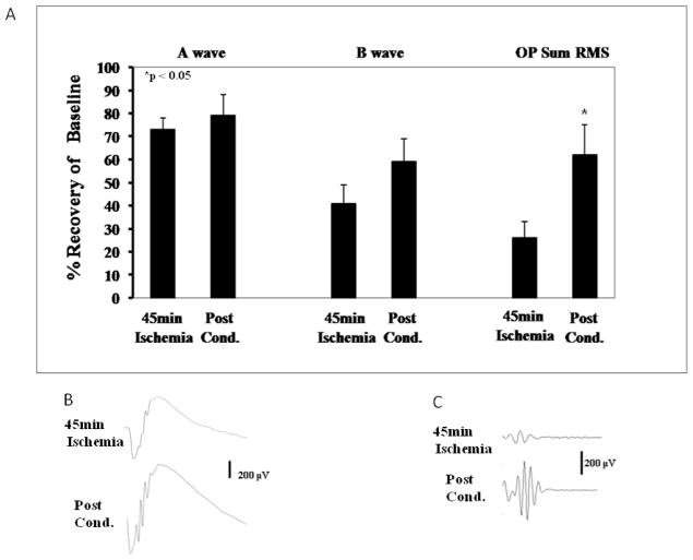
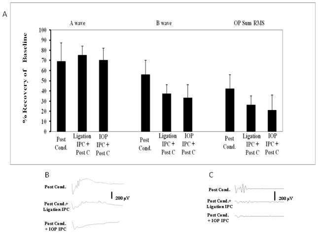
Ischemic pre-conditioning (IPC) provides neuroprotection in the rat retina from the damaging effects of severe ischemia. Recently, neuroprotection by retinal ischemic post-conditioning (Post-C), i.e., transient ischemia after more lengthy, damaging ischemia, was described, but its mechanisms are not yet known. One possible explanation of the effectiveness of Post-C is that it augments intrinsic neuroprotective mechanisms initiated during ischemia. Increasing duration of the damaging ischemic insult may therefore impact the effectiveness of Post-C. IPC, in contrast, sets in motion a series of neuroprotective events prior to the onset of ischemia. Thus, IPC and Post-C may operate by differing mechanisms. Accordingly, we examined the effect of retinal ischemic duration on post-ischemic outcome in vivo in rats after adding Post-C, and the impact of combining pre- and post-conditioning. Recovery after ischemia performed 24 h after IPC, or after Post-C performed 5 min after ischemia ended, was assessed functionally (electroretinography) and histologically at 7 days after ischemia. Durations of ischemia of 45 and 55 min were studied. Since recovery with IPC or Post-C alone, with 55 min of ischemia, did not achieve the same degree of effect (i.e., not complete recovery) exhibited in our previous studies of IPC using a different ischemia model, we also combined IPC and Post-C to test the hypothesis of the possible additive effects of the IPC and Post-C. We found that the recovery after Post-C was enhanced to a greater degree when ischemia was of longer duration. Post-C led to greater post-ischemic recovery compared to IPC. Both IPC and Post-C also attenuated structural damage to the retina. Contrary to our hypothesis, IPC and Post-C did not combine to enhance recovery after ischemia. In earlier studies, IPC attenuated post-ischemic apoptosis. To begin to examine the mechanism of Post-C, we studied its impact on apoptosis following ischemia. We examined apoptosis by determining the percentage of TUNEL-positive cells at 24 h after ischemia. Post-C attenuated apoptosis, but when combined with IPC, TUNEL was similar in the combined group to that of ischemia alone. We also examined the role of the recruitment of an inflammatory response in ischemia and Post-C. We found that inflammatory markers increased by ischemia were not altered by Post-C. We conclude that Post-C effectiveness depends upon the duration of ischemia; Post-C is not additive with IPC, and Post-C functions, in part, by preventing apoptotic damage to the inner retina. Post-C has considerable promise for clinical translation to eye diseases that cause blindness by ischemia.


Conflict of interest statement

There is no proprietary interest for any of the authors.

Figures

Figure 1
Figure 1
A. Recovery of the a- and b-waves, but not the oscillatory potentials (OPs), was relatively unchanged with Post-C (n = 9) at 7 days after ischemia as compared to the 45 min ischemia-only group (n = 8), shown at a representative flash intensity of 0.87 log cd.s/m.2 Post-C was 8 min of ischemia applied at 5 min of reperfusion after 45 min of ischemia ended; B. Representative ERG traces. RMS = root mean square amplitude of oscillatory potentials. C. Representative OP transformations.
Figure 2
Figure 2
A. Recovery of the b-wave, and the oscillatory potentials (OPs) 7 days after 55 min of ischemia was significantly enhanced by two methods of ischemic preconditioning (IPC), as compared to the 55 min ischemia group (n = 6), shown at a representative flash intensity of 0.87 log cd.s/m.2 IPC was 8 min of ischemia by elevated intraocular pressure (IOP-IPC; n = 6), or 5 min ligation of the central retinal artery (Ligation-IPC; n = 6), at 24 h prior to ischemia. B. Representative ERG traces. C. Representative OP transformations. RMS = root mean square amplitude of oscillatory potentials.
Figure 3
Figure 3
Representative histopathological images of hematoxylin and eosin-stained retinae in 5 μm thick sections for each of the experimental groups that had 55 min of ischemia. These sections were prepared from retinae removed from the rats at 7 days following ischemia. Arrows indicate layers demonstrating cell loss. Asterisks denote regions of inflammatory cell infiltration (also see Figure 5). Retinal cell layers are labeled in a normal retina in the bottom image. 55 min of ischemia resulted in retinal cell loss and disorganization of the retina, accompanied by inflammatory reaction. IPC or Post-C effectively prevented these changes, but combining IPC and Post-C produced a result resembling ischemia without IPC or Post-C.
Figure 4
Figure 4
A. Recovery of the b-wave as well as the oscillatory potentials (OPs) 7 d after ischemia was significantly enhanced with Post-C (n = 5) as compared to the 55 min ischemia group (n = 6) at a representative flash intensity of 0.87 log cd.s/m.2 Post-C was 8 min of ischemia applied 5 min after reperfusion following 55 min of ischemia. B. Representative ERG traces. C. Representative OP transformations. RMS = root mean square amplitude of oscillatory potentials.
Figure 5
Figure 5
Representative images of MCP-1, MIP-1α, and CD45-stained retinal cryosections comparing normal retinae (left column) and ischemic retinae (right column) at 7 days after ischemia. The RGC layer is at the top of each image.
Figure 6
Figure 6
A. Combining Post-C and IPC (by ligation method, “Ligation IPC,” n = 8) and by increased IOP, “IOP-IPC, n = 4) did not further enhance recovery of the a- and b-waves or OP at 7 days after 55 min of ischemia, compared to the Post-C group (n = 5) at a representative flash intensity of 0.87 log cd.s/m.2 B. Representative ERG traces. C. Representative OP transformations. RMS = root mean square amplitude of oscillatory potentials.
Figure 7
Figure 7
A. Representative fluorescent images of TUNEL in the retina at 24 h after 55 min of ischemia. Using false colors to enhance readability, red is fluorescent TUNEL, and green is DAPI. Arrows denote double-labeled (yellow) TUNEL positive retinal ganglion cells (RGC). There are numerous TUNEL cells with 55 min of ischemia, an effect significantly blunted by Post-C. IPC prior to ischemia together with Post-C following ischemia, increased TUNEL. B. Representative image of TUNEL-positive cells (green) co-localizing with Thy1 staining (red) for the 55 min of ischemia group, indicating the presence of TUNEL in retinal ganglion cells.

References

    1. Bui BV, Edmunds B, Cioffi GA, Fortune B. The gradient of retinal functional changes during acute intraocular pressure elevation. Invest Ophthalmol Vis Sci. 2005;46:202–213. - PubMed
    1. Darling CE, Jiang R, Maynard M, Whittaker P, Vinten-Johansen J, Przyklenk K. Postconditioning via stuttering reperfusion limits myocardial infarct size in rabbit hearts: role of ERK1/2. Am J Physiol. 2005;289:H1618–1626. - PubMed
    1. Dreixler JC, Barone FC, Shaikh AR, Du E, Roth S. Mitogen-activated protein kinase p38alpha and retinal ischemic preconditioning. Exp Eye Res. 2009a;89:782–790. - PMC - PubMed
    1. Dreixler JC, Hagevik S, Hemmert JW, Shaikh AR, Rosenbaum DM, Roth S. Involvement of erythropoietin in retinal ischemic preconditioning. Anesthesiology. 2009b;110:774–780. - PMC - PubMed
    1. Dreixler JC, Hemmert JW, Shenoy SK, Shen Y, Lee HT, Shaikh AR, Rosenbaum DM, Roth S. The role of Akt/protein kinase B subtypes in retinal ischemic preconditioning. Exp Eye Res. 2009c;88:512–521. - PMC - PubMed

Publication types

MeSH terms

Substances