Comparative Study
. 2010 Sep;225(1):210-8.
doi: 10.1016/j.expneurol.2010.06.017. Epub 2010 Jun 28.

Elevated tauopathy and alpha-synuclein pathology in postmortem Parkinson's disease brains with and without dementia

Affiliations
Comparative Study

Elevated tauopathy and alpha-synuclein pathology in postmortem Parkinson's disease brains with and without dementia

Jonathan Wills et al. Exp Neurol. 2010 Sep.

Abstract

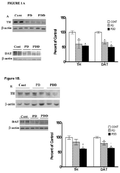
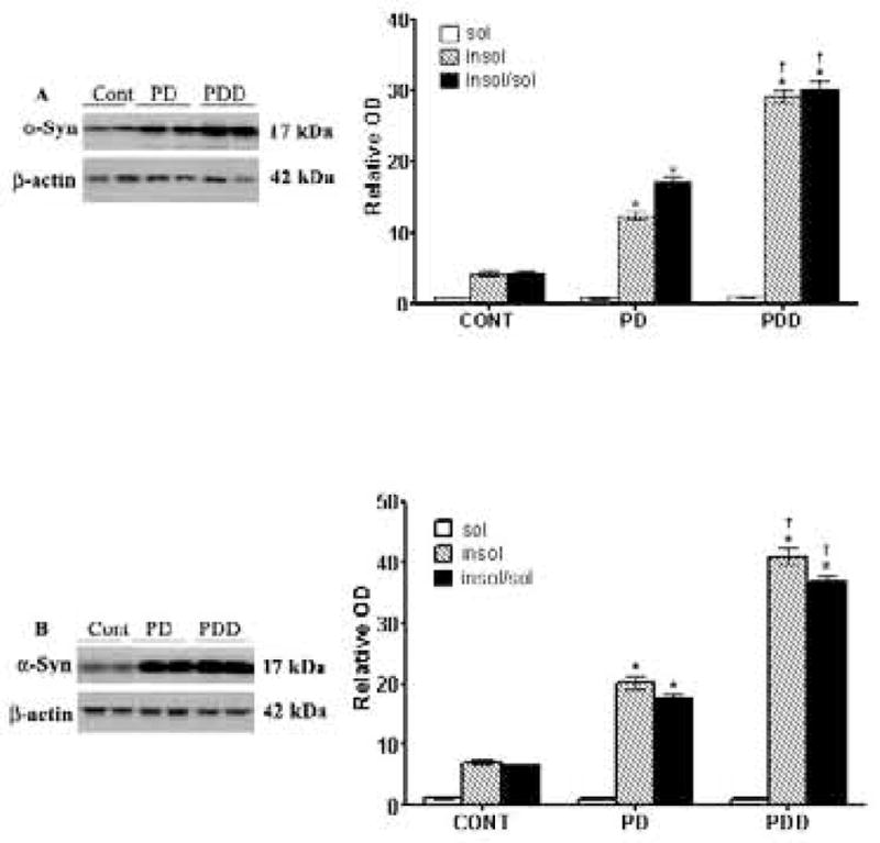
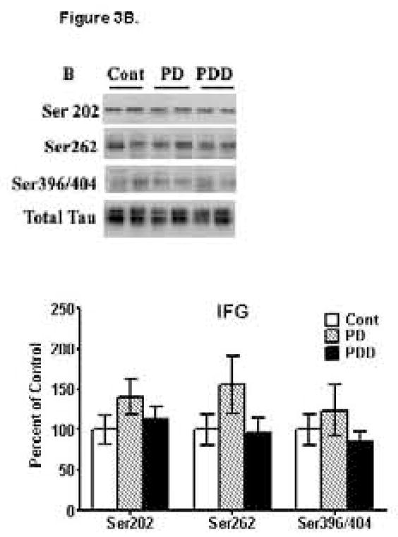
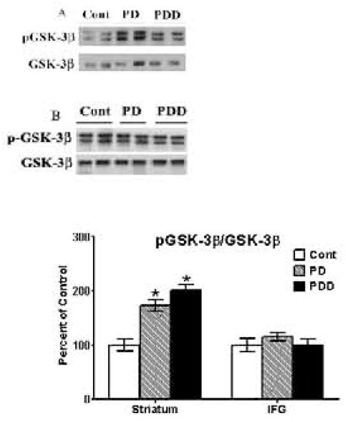
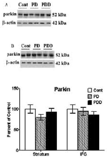
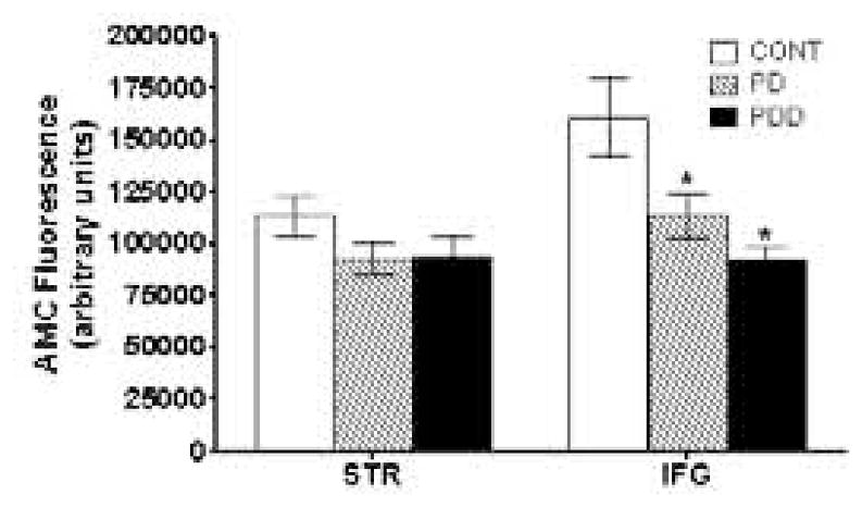
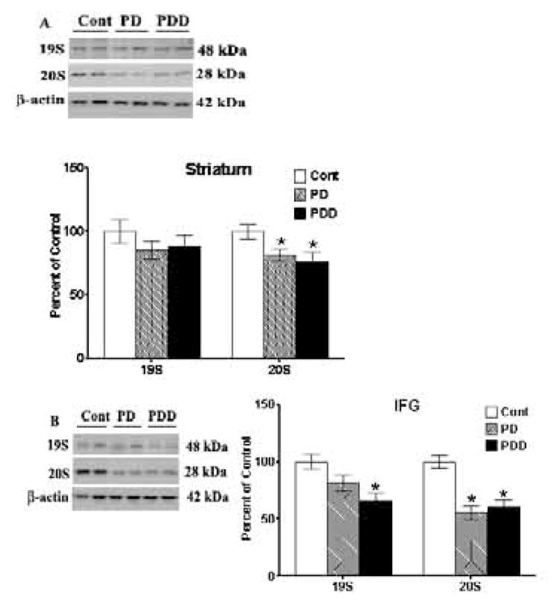
Parkinson's disease (PD), a progressive neurodegenerative disease, results in abnormal accumulation of insoluble alpha-synuclein (alpha-Syn) in dopaminergic neurons. Here we examined tauopathic changes and the alpha-Syn/p-GSK-3beta/proteasome pathway in postmortem striata and inferior frontal gyri (IFG) from patients with PD and PD with dementia (PDD). In both PD and PDD, alpha-Syn levels were high, especially the insoluble form of this protein; in PDD, insoluble alpha-Syn levels were persistently higher than PD across both brain regions. Levels of p-GSK-3beta phosphorylated at Tyr 216, which hyperphosphorylates Tau to produce toxic pathological forms of p-Tau, were higher in striata of both PD and PDD compared to controls, but were unaltered in IFG. While proteasomal activity was unchanged in striatum of PD and PDD, such activity was diminished in the IFG of both PD and PDD. A decrease in 19S subunit of the proteasomes was seen in IFG of PDD, while lower levels of 20S subunits were seen in striatum and IFG of both PD and PDD patients. Parkin levels were similar in PD and PDD, suggesting lack of involvement of this protein. Most interestingly, tauopathic changes were noted only in striatum of PD and PDD, with increased hyperphosphorylation seen at Ser262 and Ser396/404; increases in Ser202 levels were seen only in PD but not in PDD striatum. We were unable to detect any tauopathy in IFG in either PD or PDD despite increased levels of alpha-Syn, and decreased proteasomal activity, and is probably due to lack of increase in p-GSK-3beta in IFG. Unlike Alzheimer's disease where tauopathy is more globally observed in diverse brain regions, our data demonstrates restricted expression of tauopathy in brains of PD and PDD, probably limited to dopaminergic neurons of the nigrostriatal region.


Figures

Figure 1
Figure 1. Western blot analyses of tyrosine hydroxylase [TH] and dopamine transporters [DAT] in striata [A] and inferior frontal gyri [IFG] [B] from control [Cont], PD and PDD groups
Tissue lysates were prepared as described under Materials and Methods, and analyzed by Western blots using antibodies against TH and DAT. Blots are from representative experiments, while the graph is a summary of quantitation of protein levels normalized against β-actin from striata of 17 PD cases, 18 with PDD and 22 controls and while IFG are from 9 PD cases, 7 PDD and 9 controls. * denotes p<0.01 for PD and PDD groups compared to controls, while † denotes p<0.01 for PDD compared to PD.
Figure 2
Figure 2. ELISA and western blot analyses of α-synuclein [α-Syn] in striata [A] and inferior frontal gyri [IFG] [B] from control, PD and PDD groups
ELISA was conducted to measure soluble levels of α-Syn, while Western blots were conducted on insoluble α-Syn, as described in Materials and Methods. Blots are from representative experiments, while the graph is a summary of quantitation of studies from striata of 17 PD cases, 18 with PDD and 22 controls and IFG from 9 PD cases, 7 PDD and 9 controls. * denotes p<0.01 for PD and PDD groups compared to controls, while † denotes p<0.01 for PDD compared to PD.
Figure 3
Figure 3. Western blot analyses of p-Tau levels in striata [A] and inferior frontal gyri [IFG] [B] from control, PD and PDD groups
Western blot analyses of p-Tau levels were conducted using antibodies to detect hyperphosphorylation of Tau at Ser202 [CP-13 antibodies], Ser262 [anti-p-Tau-Ser262 antibodies] and Ser396/404 [PHF-1 antibodies]. Blots are from representative experiments, while the graph is a summary of quantitation of protein levels normalized against total Tau in samples, from striata of 17 PD cases, 18 with PDD and 22 controls and IFG from 9 PD cases, 7 PDD and 9 controls. * denotes p<0.01 for PD and PDD groups compared to controls.
Figure 4
Figure 4. Western blot analyses of p-GSK-3β levels in striata [A] and inferior frontal gyri [IFG] [B] from control, PD and PDD groups
Western blot analyses of p-Tau levels were conducted using antibodies to detect phosphorylation of GSK-3β at Y216. Blots are from representative experiments, while the graph is a summary of quantitation of protein levels normalized against total GSK-3β [nonphosphorylated form] in samples, from striata of 17 PD cases, 18 with PDD and 22 controls and IFG from 9 PD cases, 7 PDD and 9 controls. * denotes p<0.01 for PD and PDD groups compared to controls, while † denotes p<0.01 for PDD compared to PD.
Figure 5
Figure 5. Western blot analyses of parkin levels in striata [A] and inferior frontal gyri [IFG] [B] from control, PD and PDD groups
Western blot analyses of parkin levels were conducted using antibodies to detect parkin. Blots are from representative experiments, while the graph is a summary of quantitation of protein levels normalized against β-actin in samples, from striata of 17 PD cases, 18 with PDD and 22 controls and IFG from 9 PD cases, 7 PDD and 9 controls.
Figure 6
Figure 6. 26S Proteasome Activity in Striatum and IFG
26S proteasome activity was determined as described in Materials and Methods from striatum and IFG of PD and PDD patients, and compared with age-matched controls. Activity is reported as AMC fluorescence in arbitrary units. Statistically significant decreases in activity relative to control are indicated by an (*).
Figure 7
Figure 7. Western blot analyses of 19S and 20S levels in striata [A] and inferior frontal gyri [IFG] [B] from control, PD and PDD groups
Western blot analyses of 19S and 20S proteasomal subunits were conducted using antibodies to detect these proteins. Blots are from representative experiments, while the graph is a summary of quantitation of protein levels normalized against β-actin in samples, from striata of 17 PD cases, 18 with PDD and 22 controls and IFG from 9 PD cases, 7 PDD and 9 controls.

Comment in

References

    1. Adler CH, Hentz JG, Joyce JN, Beach T, Caviness JN. Motor Impairment in Normal Aging, Clinically Possible Parkinson’s Disease, and Clinically Probable Parkinson’s Disease: Longitudinal Evaluation of a Cohort of Prospective Brain Donors. Parkinsonism and Related Disorders. 2002;9:103–110. - PubMed
    1. Arai Y, Yamazaki M, Mori O, Muramatsu H, Asano G, Katayama Y. Alpha-synuclein-positive structures in cases with sporadic Alzheimer’s disease: morphology and its relationship to tau aggregation. Brain Res. 2001;888:287–296. - PubMed
    1. Baum L, Seger R, Woodgett J, Kawabata S, Maruyama K, Koyama M, Silver J, Saitoh T. Overexpressed tau protein in cultured cells is phosphorylated without formation of PHF: implication of phosphoprotein phosphatase involvement. Mol Brain Res. 1995;34:1–17. - PubMed
    1. Beyer K, Domingo-Sàbat M, Ariza A. Molecular pathology of lewy body diseases. Int J Mol Sci. 2009;10:724–45. - PMC - PubMed
    1. Burns J, Galvin JE, Roe CM, Morris JC, McKeel DW. The pathology of the substantia nigra in Alzheimer disease with extrapyramidal signs. Neurology. 2005;64:1397–403. - PubMed

Publication types

MeSH terms