. 2010 Sep 3;401(5):985-95.
doi: 10.1016/j.jmb.2010.06.042. Epub 2010 Jun 26.

Disulfide bond stabilization of the hexameric capsomer of human immunodeficiency virus

Affiliations

Disulfide bond stabilization of the hexameric capsomer of human immunodeficiency virus

Owen Pornillos et al. J Mol Biol. .

Abstract

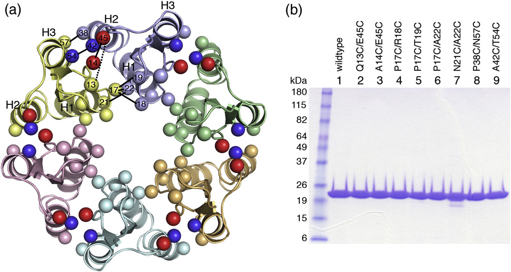
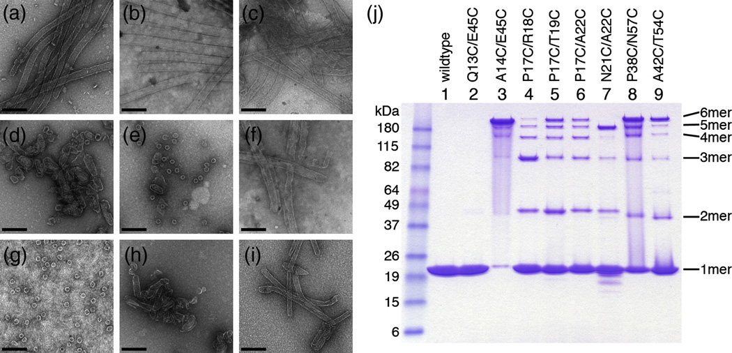
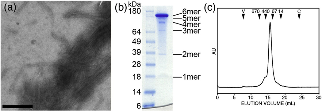
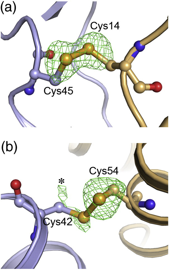
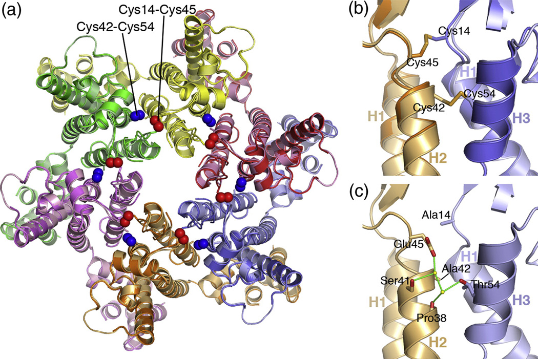
The human immunodeficiency virus type 1 capsid is modeled as a fullerene cone that is composed of approximately 250 hexamers and 12 pentamers of the viral CA protein. Structures of CA hexamers have been difficult to obtain because the hexamer-stabilizing interactions are inherently weak, and CA tends to spontaneously assemble into capsid-like particles. Here, we describe a two-step biochemical strategy to obtain soluble CA hexamers for crystallization. First, the hexamer was stabilized by engineering disulfide cross-links (either A14C/E45C or A42C/T54C) between the N-terminal domains of adjacent subunits. Second, the cross-linked hexamers were prevented from polymerizing further into hyperstable capsid-like structures by mutations (W184A and M185A) that interfered with dimeric association between the C-terminal domains that link adjacent hexamers. The structures of two different cross-linked CA hexamers were nearly identical, and we combined the non-mutated portions of the structures to generate an atomic resolution model for the native hexamer. This hybrid approach for structure determination should be applicable to other viral capsomers and protein-protein complexes in general.


Figures

Figure 1
Figure 1
Model-based design of disulfide-stabilized HIV-1 CA hexamers. (A) Top view ribbons representation of the 18-helix barrel comprising the NTD-NTD hexamerization interactions, as seen in the cryoEM model (3dik). Each subunit is colored differently. The locations of mutated residues are indicated by spheres, and are labeled within a single interface (between yellow and blue subunits). Pairs are connected by black lines (dashed for the Q13/E45 negative control). The A14/E45 and A42/T54 pairs are colored red and blue, respectively. (B) SDS-PAGE profile of purified HIV-1 CA double cysteine mutants. Ten µg protein was loaded in each lane. Molecular weight markers are labeled on the left.
Figure 2
Figure 2
Assembly and crosslinking of double cysteine CA mutants. (A–I) Representative negatively stained EM images of in vitro assembled (A) Wildtype HIV-1 CA, (B) Q13C/E45C, (C) A14C/E45C, (D) P17C/R18C, (E) P17C/T19C, (F) P17C/A22C, (G) N21C/A22C, (H) P38C/N57C, and (I) A42C/T54C. The assembly buffer contained 10 mg/mL protein in 50 mM Tris, pH 8, 1 M NaCl, 20 mM βME. Scale bar = 200 nm. (J) Non-reducing SDS-PAGE profiles of the assembly reactions. Molecular weight markers are labeled on the left, and the positions of crosslinked n-mers are indicated on the right.
Figure 3
Figure 3
Assembly and analysis of A14C/E45C/W184A/M185A (CC1 construct). (A) Negatively-stained EM image of tubes assembled by dialyzing 30 mg/mL protein in 50 mM Tris, pH 8, 1 M NaCl, 2 mM βME. Scale bar = 500 nm. (B) Non-reducing SDS-PAGE profile of crosslinked soluble CC1 hexamers. Molecular weight markers are labeled on the left, and the expected positions of crosslinked n-mers are indicated on the right. (C) Size exclusion chromatographic profile of soluble crosslinked CC1. The elution volumes of protein standards are indicated by black arrowheads (numbers indicate molecular weight in kDa, V = void volume, C = column volume). The A42C/T54C/W184A/M185A mutant (CC2 construct) behaved similarly, but only crosslinked into hexamers with ~50% efficiency (data not shown).
Figure 4
Figure 4
Conformations of the engineered disulfide bonds. (A) Cys14 and Cys45 in the CC1 construct, shown in ball-and-stick representation, with the rest of the structure in ribbons. Green mesh show omit mFo-DFc densities for the sulfur atoms contoured at 3σ. The omit map was obtained by setting the sulfur occupancies to zero, introducing random shifts to the remaining atoms (mean displacement = 0.5 Å), and refining the resulting model with simulated annealing. Note that the sulfur Cys14 and Cys45 sulfur density peaks have equal magnitude and are ~100% oxidized. (B) Disulfide-bonded conformation of Cys42 and Cys54 in the CC2 construct. Note that the sulfur density for Cys54 is clear, whereas Cys42 is weaker and more diffuse. The asterisk indicates density ascribed to an alternative rotamer configuration for Cys42, which is not disulfide-bonded.
Figure 5
Figure 5
Composite structure of the HIV-1 CA hexamer. (A) Superposition of the CC1 and CC2 hexamer structures viewed as would be seen from the outside of the capsid. Each subunit is colored differently, with CC1 is brighter shades and CC2 in muted shades. The engineered Cys14/Cys45 and Cys42/Cys54 disulfide bonds are indicated by the red and blue spheres, respectively. (B) Close-up side view of the superposition in the vicinity of the engineered disulfides. (C) View of the composite hexamer interface model, in the same orientation as (B). Sidechains for Ala14, Ala42, Glu45, and Thr54 are modeled in their native conformations. A subset of ordered water molecules within the interface are shown as yellow spheres. Putative hydrogen bonds are shown by green lines.
Figure 6
Figure 6
Stereoview of the β-hairpin region between two adjacent subunits. Details of the hydrophilic interactions surrounding the Pro1-Asp51 salt bridge (asterisk) are shown for one subunit, as seen in the composite hexamer model. Similar arrangements were observed each of the crystallographically independent CC1 and CC2 hexamer structures. Note that there are essentially no intermolecular interactions involving either the salt bridge or strand regions of the β-hairpin.

References

    1. Ganser-Pornillos BK, Yeager M, Sundquist WI. The structural biology of HIV assembly. Curr Opin Struct Biol. 2008;18:203–217. - PMC - PubMed
    1. Jin Z, Jin L, Peterson DL, Lawson CL. Model for lentivirus capsid core assembly based on crystal dimers of EIAV p26. J Mol Biol. 1999;286:83–93. - PubMed
    1. Li S, Hill CP, Sundquist WI, Finch JT. Image reconstructions of helical assemblies of the HIV-1 CA protein. Nature. 2000;407:409–413. - PubMed
    1. Ganser BK, Li S, Klishko VY, Finch JT, Sundquist WI. Assembly and analysis of conical models for the HIV-1 core. Science. 1999;283:80–83. - PubMed
    1. Mortuza GB, Haire LF, Stevens A, Smerdon SJ, Stoye JP, Taylor IA. High-resolution structure of a retroviral capsid hexameric amino-terminal domain. Nature. 2004;431:481–485. - PubMed

Publication types

Associated data

LinkOut - more resources