. 2010 Aug 20;401(3):503-17.
doi: 10.1016/j.jmb.2010.06.023. Epub 2010 Jun 18.

Abeta(1-40) forms five distinct amyloid structures whose beta-sheet contents and fibril stabilities are correlated

Affiliations

Abeta(1-40) forms five distinct amyloid structures whose beta-sheet contents and fibril stabilities are correlated

Ravindra Kodali et al. J Mol Biol. .

Abstract

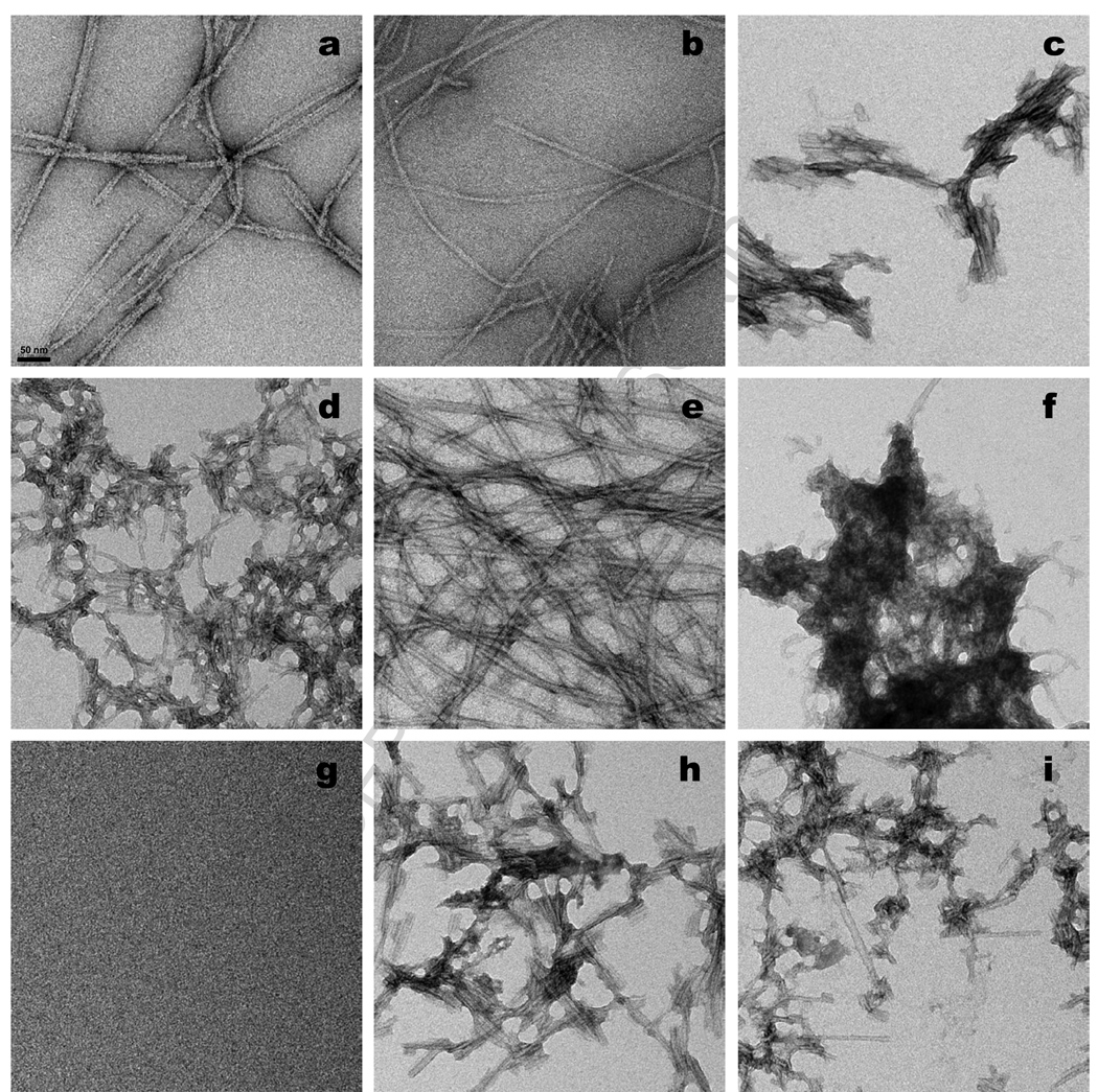
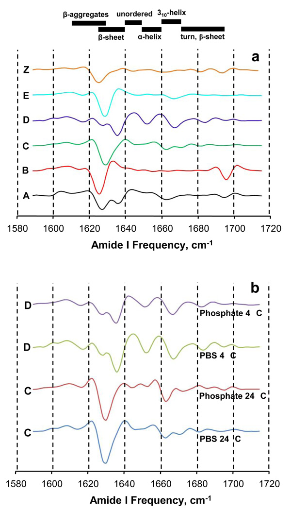
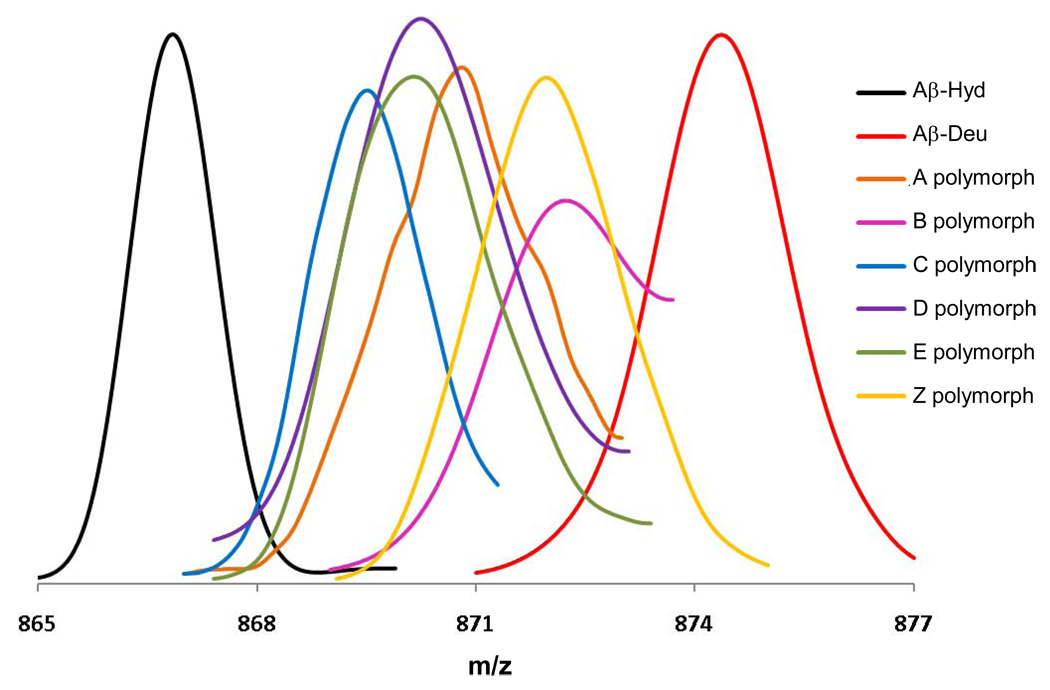
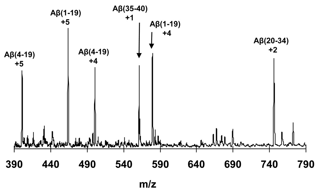
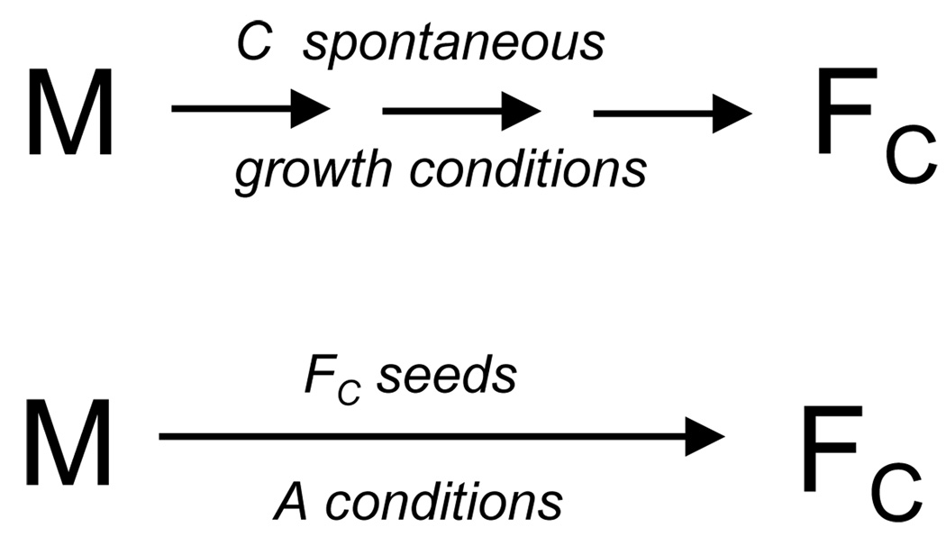
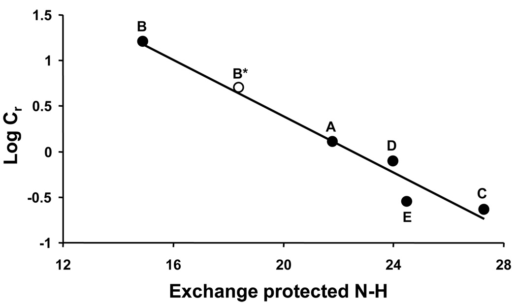
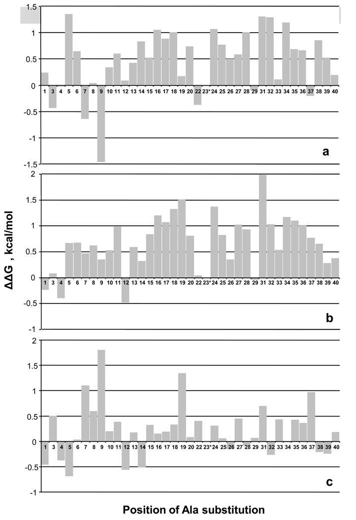
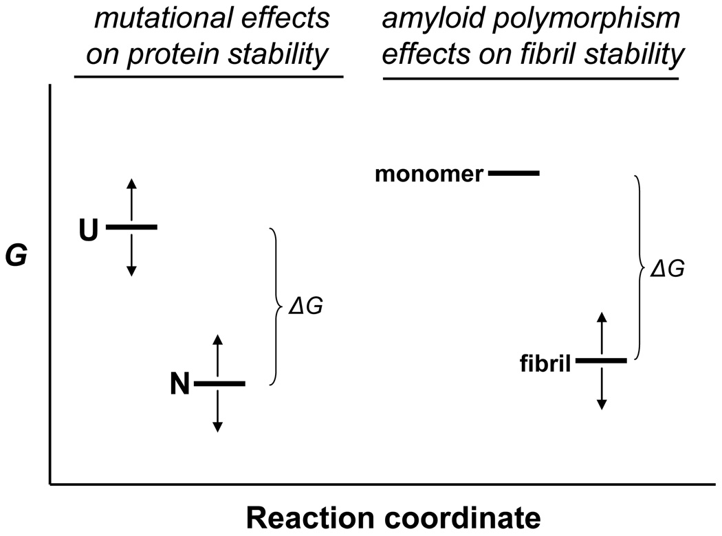
The ability of a single polypeptide sequence to grow into multiple stable amyloid fibrils sets these aggregates apart from most native globular proteins. The existence of multiple amyloid forms is the basis for strain effects in yeast prion biology, and might contribute to variations in Alzheimer's disease pathology. However, the structural basis for amyloid polymorphism is poorly understood. We report here five structurally distinct fibrillar aggregates of the Alzheimer's plaque peptide Abeta(1-40), as well as a non-fibrillar aggregate induced by Zn(2+). Each of these conformational forms exhibits a unique profile of physical properties, and all the fibrillar forms breed true in elongation reactions under a common set of growth conditions. Consistent with their defining cross-beta structure, we find that in this series the amyloid fibrils containing more extensive beta-sheet exhibit greater stability. At the same time, side chain packing outside of the beta-sheet regions contributes to stability, and to differences of stability between polymorphic forms. Stability comparison is facilitated by the unique feature that the free energy of the monomer (equivalent to the unfolded state in a protein folding reaction) does not vary, and hence can be ignored, in the comparison of DeltaG degrees of elongation values for each polymorphic fibril obtained under a single set of conditions.


Figures

Figure 1
Figure 1
Amino acid sequence of human Aβ(1–40).
Figure 2
Figure 2
Negative stained electron micrographs of samples freshly harvested from aggregation reactions. (a) polymorph A; (b) polymorph B; (c) polymorph C made in PBS; (d) polymorph D made in PBS; (e) polymorph E; (f) polymorph Z; (g) mock staining of grid treated with ZnCl2 in PBS; (h) polymorph C made in phosphate buffer; (i) polymorph D made in phosphate buffer. Scale bar, 50 nm.
Figure 3
Figure 3
Second derivative FTIR spectra of various aggregated polymorphic forms of Aβ(1–40). Secondary structure frequency ranges at the top of the figure are estimates from reference . (a) Comparison of the six polymorphic forms described herein. (b) Comparisons of the two sets of sibling fibrils for the C and D polymorphs, each prepared in either PBS or phosphate buffer.
Figure 4
Figure 4
Electrospray mass spectrograms of the +5 charge state of the polymorphic Aβ(1–40) aggregate preparations described in this paper. “Aβ-Hyd” and “Aβ-Deu” are the Aβ(1–40) A polymorph fibrils grown in either H2O or D2O. Polymorphic fibrils (and the Zn induced aggregate “Z”) were grown in H2O, harvested, and subjected to D2O exchange for 24 hrs. All were analyzed by in-line H/D exchange as described, in which fibrils are dissolved in-line and streamed into the MS. Small peaks appearing at the “fully exchanged” m/z value at the “Aβ-Deu” position, which appear to be due to equilibrium exchange of deuterated monomeric Aβ(1–40) into fibrils during the exchange incubation , are not shown.
Figure 5
Figure 5
Mass spectrogram resulting from dissolution of a suspension of A type Aβ(1–40) H-fibrils with pepsin in aqueous formic acid in the mixing stream of a T-tube front end of the mass spectrometer, showing the major charge state species of the resulting pepsin digestion fragments.
Figure 6
Figure 6
Schematic summary of seeding experiments. In this example, monomeric Aβ(1–40) “M” is incubated under the solution conditions (agitation, PBS, 24 °C) that generate the type C polymorph. The type C fibrils (“FC”) are isolated, then used to seed a solution of monomer incubated under the normal conditions (PBS, 37 °C, no agitation) for obtaining the type A polymorph. If fibril structure is, as expected, controlled by the structure of the seed and not by the incubation conditions, the product fibrils should also be of the C type. Since the elongation equilibrium is achieved in this case under A conditions, however, the Cr and derived ΔG°elong should reflect this.
Figure 7
Figure 7
Correlation of fibril stability with β-sheet content. Cr values were obtained from seeded elongation fibril reactions conducted in phosphate buffer, where seeding was with different fibril polymorphs, as shown in a general way in Figure 6 (Table 1). Log Cr (●) is plotted against corrected HX-MS values of each fibril product. Slope = − 0.1548, R2 = 0.9598. The open circle (○) represents the data for the product (“B*”) of elongation in PBS at 37 °C of Aβ(1–40) monomers seeded with B fibrils. This data point was not used in the linear fit.
Figure 8
Figure 8
Impact of Ala mutations on Aβ(1–40) amyloid fibril stability. Monomeric Aβ(1–40) Ala point mutants were subjected to spontaneous growth conditions and the Cr values assessed after ThT signal development reached a plateau. ΔG°elong values were calculated from the Cr values . Bar graphs represent pairwise comparisons, with positive ΔΔG values indicating lower stability. (a) ΔG°WT − ΔG°Ala for fibrils grown under C conditions; positive values indicate the Ala mutation decreases fibril stability. (b) ΔG°WT − ΔG°Ala for fibrils grown under A conditions; positive values indicate the Ala mutation decreases fibril stability. Data from reference . (c) ΔG °A − ΔG°C for each Ala point mutant. Positive values indicate that the Ala mutation is more destabilizing of A fibrils than of C fibrils, or, in other words, the Ala mutation is better accommodated into C fibrils than into A fibrils.
Figure 9
Figure 9
Interpretation of free energy changes in protein transformations. In mutational analyses of protein folding reactions, mutations can modify the free energy (G) of both the unfolded state (U) and the folded, native state (N), as indicated by the arrows. This means that the physically measurable parameter, ΔG° - the free energy change on folding - cannot be rigorously ascribed to a change in the free energy of the folded state. In contrast, in the comparison of free energy changes of fibril elongation for different polymorphic fibrils from the same amino acid sequence, as determined in seeded elongation of these polymorphs under identical solution conditions, the difference in measured ΔG° values between polymorphic fibrils must be identical to the difference in free energies between the fibrils themselves, since the G of the monomer does not change under the standard elongation conditions used.

References

    1. Martin JB. Molecular basis of the neurodegenerative disorders. N Engl J Med. 1999;340:1970–1980. [published erratum appears in N Engl J Med 1999 Oct 28;341(18):1407] - PubMed
    1. Merlini G, Bellotti V. Molecular mechanisms of amyloidosis. N Engl J Med. 2003;349:583–596. - PubMed
    1. Wetzel R, Shivaprasad S, Williams AD. Plasticity of amyloid fibrils. Biochemistry. 2007;46:1–10. - PMC - PubMed
    1. Serpell LC, Fraser PE, Sunde M. X-ray fiber diffraction of amyloid fibrils. Meth Enzymol. 1999;309:526–536. - PubMed
    1. Luhrs T, Ritter C, Adrian M, Riek-Loher D, Bohrmann B, Dobeli H, Schubert D, Riek R. 3D structure of Alzheimer's amyloid-beta(1–42) fibrils. Proc Natl Acad Sci U S A. 2005;102:17342–17347. - PMC - PubMed

Publication types

LinkOut - more resources