Oxygen and oxygenation in stem-cell therapy for myocardial infarction
- PMID: 20600148
- PMCID: PMC3114881
- DOI: 10.1016/j.lfs.2010.06.013
Oxygen and oxygenation in stem-cell therapy for myocardial infarction
Abstract
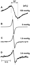
Myocardial infarction (MI) is caused by deprivation of oxygen and nutrients to the cardiac tissue due to blockade of coronary artery. It is a major contributor to chronic heart disease, a leading cause of mortality in the modern world. Oxygen is required to meet the constant energy demands for heart contractility, and also plays an important role in the regulation of heart function. However, reoxygenation of the ischemic myocardium upon restoration of blood flow may lead to further injury. Controlled oxygen delivery during reperfusion has been advocated to prevent this consequence. Monitoring the myocardial oxygen concentration would play a vital role in understanding the pathological changes in the ischemic heart following myocardial infarction. During the last two decades, several new techniques have become available to monitor myocardial oxygen concentration in vivo. Electron paramagnetic resonance (EPR) oximetry would appear to be the most promising and reliable of these techniques. EPR utilizes crystalline probes which yield a single sharp line, the width of which is highly sensitive to oxygen tension. Decreased oxygen tension results in a sharpening of the EPR spectrum, while an increase results in widening. In our recent studies, we have used EPR oximetry as a valuable tool to monitor myocardial oxygenation for several applications like ischemia-reperfusion injury, stem-cell therapy and hyperbaric oxygen therapy. The results obtained from these studies have demonstrated the importance of tissue oxygen in the application of stem-cell therapy to treat ischemic heart tissues. These results have been summarized in this review article.
Copyright (c) 2010 Elsevier Inc. All rights reserved.
Figures


Similar articles
-
Direct and Repeated Measurement of Heart and Brain Oxygenation Using In Vivo EPR Oximetry.Methods Enzymol. 2015;564:529-52. doi: 10.1016/bs.mie.2015.06.023. Epub 2015 Jul 21. Methods Enzymol. 2015. PMID: 26477264
-
Myocardial oxygenation and functional recovery in infarct rat hearts transplanted with mesenchymal stem cells.Am J Physiol Heart Circ Physiol. 2009 May;296(5):H1263-73. doi: 10.1152/ajpheart.01311.2008. Epub 2009 Mar 13. Am J Physiol Heart Circ Physiol. 2009. PMID: 19286938 Free PMC article.
-
EPR oximetry in the beating heart: myocardial oxygen consumption rate as an index of postischemic recovery.Magn Reson Med. 2004 Apr;51(4):835-42. doi: 10.1002/mrm.20000. Magn Reson Med. 2004. PMID: 15065258
-
Beyond Reperfusion: Acute Ventricular Unloading and Cardioprotection During Myocardial Infarction.J Cardiovasc Transl Res. 2019 Apr;12(2):95-106. doi: 10.1007/s12265-019-9863-z. Epub 2019 Jan 22. J Cardiovasc Transl Res. 2019. PMID: 30671717 Free PMC article. Review.
-
Role of oxygen in postischemic myocardial injury.Antioxid Redox Signal. 2007 Aug;9(8):1193-206. doi: 10.1089/ars.2007.1636. Antioxid Redox Signal. 2007. PMID: 17571958 Review.
Cited by
-
Optimization of the cardiovascular therapeutic properties of mesenchymal stromal/stem cells-taking the next step.Stem Cell Rev Rep. 2013 Jun;9(3):281-302. doi: 10.1007/s12015-012-9366-7. Stem Cell Rev Rep. 2013. PMID: 22529015 Review.
-
Hypoxic preconditioning induces the expression of prosurvival and proangiogenic markers in mesenchymal stem cells.Am J Physiol Cell Physiol. 2010 Dec;299(6):C1562-70. doi: 10.1152/ajpcell.00221.2010. Epub 2010 Sep 22. Am J Physiol Cell Physiol. 2010. PMID: 20861473 Free PMC article.
-
Hypoxic preconditioning effect on stromal cells derived factor-1 and C-X-C chemokine receptor type 4 expression in Wistar rat's (Rattus norvegicus) bone marrow mesenchymal stem cells (in vitro study).Vet World. 2018 Jul;11(7):965-970. doi: 10.14202/vetworld.2018.965-970. Epub 2018 Jul 19. Vet World. 2018. PMID: 30147267 Free PMC article.
-
Selective inhibition of hypoxia-inducible factor 1α ameliorates adipose tissue dysfunction.Mol Cell Biol. 2013 Mar;33(5):904-17. doi: 10.1128/MCB.00951-12. Epub 2012 Dec 17. Mol Cell Biol. 2013. PMID: 23249949 Free PMC article.
-
Hypoxic preconditioning improves survival of cardiac progenitor cells: role of stromal cell derived factor-1α-CXCR4 axis.PLoS One. 2012;7(7):e37948. doi: 10.1371/journal.pone.0037948. Epub 2012 Jul 18. PLoS One. 2012. PMID: 22815687 Free PMC article.
References
-
- Agbulut O, Vandervelde S, Al Attar N, Larghero J, Ghostine S, Leobon B, Robidel E, Borsani P, Le Lorc'h M, Bissery A, Chomienne C, Bruneval P, Marolleau JP, Vilquin JT, Hagege A, Samuel JL, Menasche P. Comparison of human skeletal myoblasts and bone marrow-derived CD133+ progenitors for the repair of infarcted myocardium. Journal of the American College of Cardiology. 2004;44(2):458–463. - PubMed
-
- Ambrosio G, Jacobus WE, Bergman CA, Weisman HF, Becker LC. Preserved high energy phosphate metabolic reserve in globally “stunned” hearts despite reduction of basal ATP content and contractility. Journal of Molecular and Cellular Cardiology. 1987;19(10):953–964. - PubMed
-
- Arnesen H, Lunde K, Aakhus S, Forfang K. Cell therapy in myocardial infarction. Lancet. 2007;369(9580):2142–2143. - PubMed
-
- Bolli R, Marban E. Molecular and cellular mechanisms of myocardial stunning. Physiological Reviews. 1999;79(2):609–634. - PubMed
Publication types
MeSH terms
Grants and funding
LinkOut - more resources
Full Text Sources
Other Literature Sources
Medical

