

Impaired fixation to eyes following amygdala damage arises from abnormal bottom-up attention
- PMID: 20600184
- PMCID: PMC2949539
- DOI: 10.1016/j.neuropsychologia.2010.06.025
Impaired fixation to eyes following amygdala damage arises from abnormal bottom-up attention
Abstract
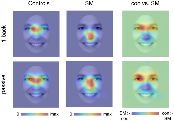
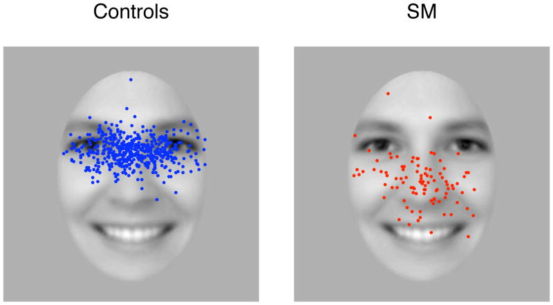
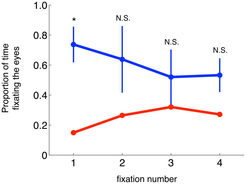
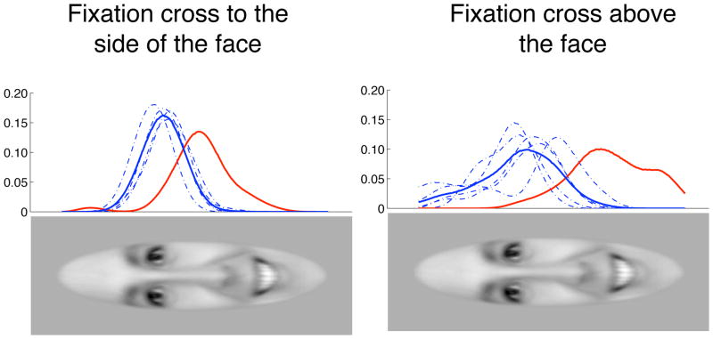
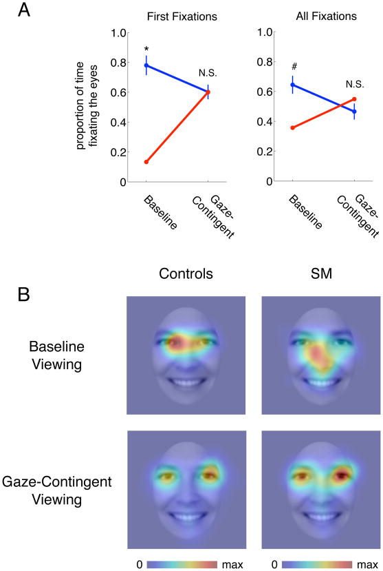
SM is a patient with complete bilateral amygdala lesions who fails to fixate the eyes in faces and is consequently impaired in recognizing fear (Adolphs et al., 2005). Here we first replicated earlier findings in SM of reduced gaze to the eyes when seen in whole faces. Examination of the time course of fixations revealed that SM's reduced eye contact is particular pronounced in the first fixation to the face, and less abnormal in subsequent fixations. In a second set of experiments, we used a gaze-contingent presentation of faces with real time eye tracking, wherein only a small region of the face is made visible at the center of gaze. In essence, viewers explore the face by moving a small searchlight over the face with their gaze. Under such viewing conditions, SM's fixations to eye region of faces became entirely normalized. We suggest that this effect arises from the absence of bottom-up effects due to the facial features, allowing gaze location to be driven entirely by top-down control. Together with SM's failure to fixate the eyes in whole faces primarily at the very first saccade, the findings suggest that the saliency of the eyes normally attract our gaze in an amygdala-dependent manner. Impaired eye gaze is also a prominent feature of several psychiatric illnesses in which the amygdala has been hypothesized to be dysfunctional, and our findings and experimental manipulation may hold promise for interventions in such populations, including autism and fragile X syndrome.
Copyright © 2010 Elsevier Ltd. All rights reserved.
Figures

Republished in
-
Reprint of: Impaired fixation to eyes following amygdala damage arises from abnormal bottom-up attention.Neuropsychologia. 2011 Mar;49(4):589-95. doi: 10.1016/j.neuropsychologia.2011.02.026. Neuropsychologia. 2011. PMID: 21414461 Free PMC article.
References
-
- Adolphs R, Tranel D, Damasio H, Damasio A. Impaired recognition of emotion in facial expressions following bilateral damage to the human amygdala. Nature. 1994;372:669–672. - PubMed
-
- Adolphs R, Gosselin F, Buchanan TW, Tranel D, Schyns P, Damasio AR. A mechanism for impaired fear recognition after amygdala damage. Nature. 2005;433:68–72. - PubMed
-
- Adolphs R, Spezio M. Role of the amygdala in processing visual social stimuli. Prog Brain Res. 2006;156:363–378. - PubMed
-
- Adolphs R, Tranel D, Hamann S, Young AW, Calder AJ, Phelps EA, Anderson A, Lee GP, Damasio AR. Recognition of facial emotion in nine individuals with bilateral amygdala damage. Neuropsychologia. 1999;37:1111–1117. - PubMed
-
- Brainard DH. The Psychophysics Toolbox. Spat Vis. 1997;10:433–436. - PubMed
Publication types
MeSH terms
Grants and funding
LinkOut - more resources
Full Text Sources
Other Literature Sources
Medical
