Comparative Study
. 2010 Dec;31(6):738-46.
doi: 10.1016/j.neuro.2010.06.007. Epub 2010 Jul 1.

Soman increases neuronal COX-2 levels: possible link between seizures and protracted neuronal damage

Affiliations
Comparative Study

Soman increases neuronal COX-2 levels: possible link between seizures and protracted neuronal damage

Mariana Angoa-Pérez et al. Neurotoxicology. 2010 Dec.

Abstract

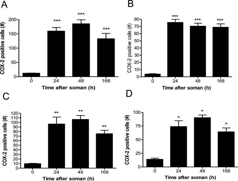
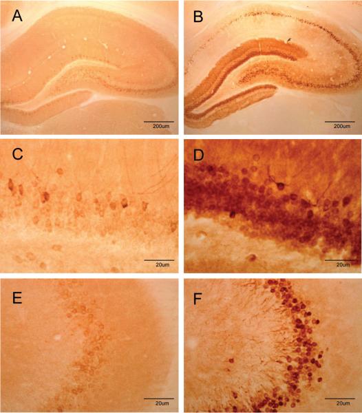
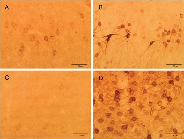
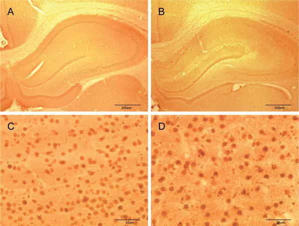
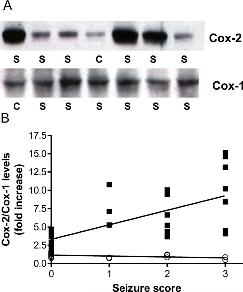
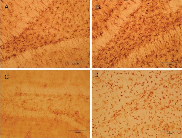
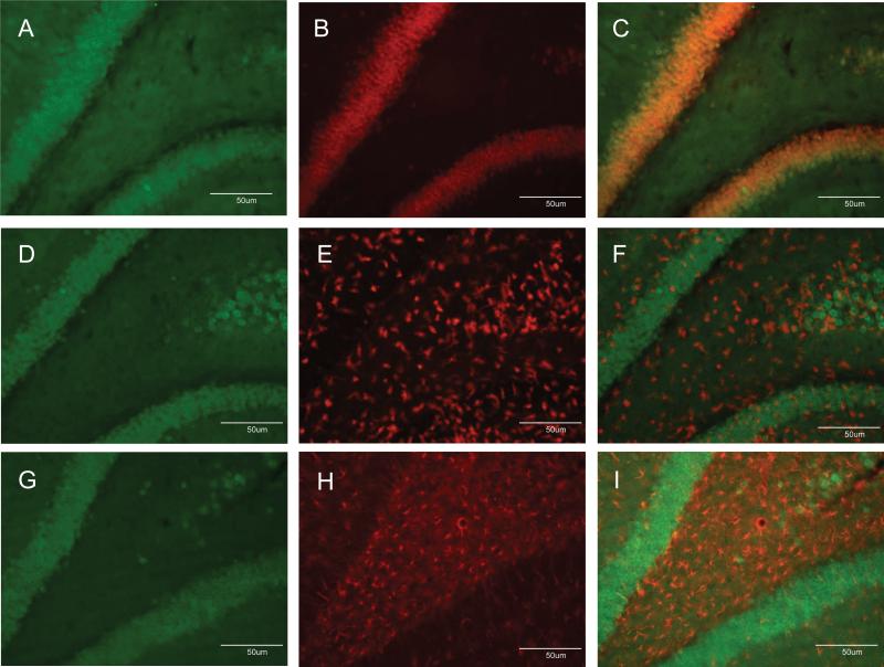
Nerve agent-induced seizures cause neuronal damage in brain limbic and cortical circuits leading to persistent behavioral and cognitive deficits. Without aggressive anticholinergic and benzodiazepine therapy, seizures can be prolonged and neuronal damage progresses for extended periods of time. The objective of this study was to determine the effects of the nerve agent soman on expression of cyclooxygenase-2 (COX-2), the initial enzyme in the biosynthetic pathway of the proinflammatory prostaglandins and a factor that has been implicated in seizure initiation and propagation. Rats were exposed to a toxic dose of soman and scored behaviorally for seizure intensity. Expression of COX-2 was determined throughout brain from 4h to 7 days after exposure by immunohistochemistry and immunoblotting. Microglial activation and astrogliosis were assessed microscopically over the same time-course. Soman increased COX-2 expression in brain regions known to be damaged by nerve agents (e.g., hippocampus, amygdala, piriform cortex and thalamus). COX-2 expression was induced in neurons, and not in microglia or astrocytes, and remained elevated through 7 days. The magnitude of COX-2 induction was correlated with seizure intensity. COX-1 expression was not changed by soman. Increased expression of neuronal COX-2 by soman is a late-developing response relative to other signs of acute physiological distress caused by nerve agents. COX-2-mediated production of prostaglandins is a consequence of the seizure-induced neuronal damage, even after survival of the initial cholinergic crisis is assured. COX-2 inhibitors should be considered as adjunct therapy in nerve agent poisoning to minimize nerve agent-induced seizure activity.


Figures

Fig 1
Fig 1
Soman increases COX-2 expression in brain. Rats (N= 5 per group) were treated with soman and brains were processed for immunohistochemistry at the indicated times after treatment using antibodies against COX-2 as described in Materials and Methods. COX-2 positive cells were counted in dentate gyrus (A), CA3 (B), amygdala (C) and piriform cortex (D). Very few COX-2 positive cells were seen from 0-12 hours after soman so these data are not shown. Results are presented as number of COX-2 positive cells in each brain region ± SEM. *p < 0.05; **p < 0.01; ***p < 0.001, Tukey's multiple comparison test by comparison to controls.
Fig 2
Fig 2
Soman increases COX-2 expression in the hippocampus. Rats (N= 5 per group) were treated with soman and brains were prepared for immunohistochemistry 48 hr after treatment using antibodies against COX-2. Few COX-2 positive cells are seen in controls (A) whereas the dentate gyrus, CA1 and CA3 are heavily labeled with COX-2 after soman treatment (B). Higher magnification images of the dentate gyrus shows sparse labeling of cells in controls (C) and densely packed COX-2-positive cells in soman-treated rats (D). Similarly, higher magnification images from the CA3 region of controls shows few COX-2-positive cells in controls (E) whereas soman treatment causes a large increase in COX-2 levels (F).
Fig 3
Fig 3
Soman increases COX-2 expression in the piriform cortex and amygdala. Rats (N= 5 per group) were treated with soman and brains were prepared for immunohistochemistry 48 hr after treatment using antibodies against COX-2. Few COX-2 positive cells are seen in the piriform cortex of controls (A) whereas numerous cells and processes are COX-2 positive after soman treatment (B). Soman also leads to large increases in COX-2 expression in the amygdala (D) as compared to controls (C).
Fig 4
Fig 4
Soman does not change COX-1 expression in brain. Rats (N= 5 per group) were treated with soman and brains were processed for immunohistochemistry 48 hr after treatment using antibodies against COX-1 as described in Materials and Methods. Few COX-1 positive cells are seen in the hippocampus of controls (A) or soman (B) treated rats at low power magnification. Images collected from the amygdala at higher magnification showed numerous COX-1 positive cells in controls (C) but soman treatment (D) did not increase the number of these cells.
Fig 5
Fig 5
Soman-induced increases in COX-2 expression are correlated with seizure intensity. Rats (N= 5-8 per group) were treated with soman and scored behaviorally for seizure intensity 1 hr after treatment as described in Materials and Methods. At 48 hr after soman, brains were processed for immunoblotting for COX-2 and COX-1 in the hippocampus. Panel A shows immunoblotting results for COX-2 and COX-1 from controls (C) and soman treated rats (S). The fold-increase in COX-2 (■) and COX-1 (○) expression caused by soman was plotted versus seizure intensity in panel B. The relationship between changes in COX-2 levels and seizure intensity was significant (r=0.73, p < 0.0001). COX-1 levels were not changed by soman treatment.
Fig 6
Fig 6
Soman causes gliosis in hippocampus. Rats were treated with soman and brains were processed for immunohistochemistry 48 hr after treatment. Astrocytes were labeled in control (A) or soman-treated rats (B) using antibodies against GFAP. Activated microglia were labeled in control (C) or soman-treated rats (D) using ILB4 staining.
Fig 7
Fig 7
Soman increases COX-2 expression solely in neurons. Rats were treated with soman and brains were processed after 48 hr for double labeling fluorescence immunohistochemistry as described in Materials and Methods. COX-2-positive cells were labeled with Alexa 488-conjugated antibodies (green fluorescence, panels A, D, G). Neurons were identified using antibodies against NeuN and Alexa-555-conjugated secondary antibodies (orange fluorescence, panel B). Activated microglial were identified using Alexa-568-conjugated ILB4 (red fluorescence, panel E). Astrocytes were identified using antibodies against GFAP and Alexa-647-conjugated secondary antibodies (red fluorescence, panel H). Merged images in panels C, F and I indicate that COX-2 labeling overlapped with neurons and not with microglia or astrocytes.

References

    1. Baille V, Clarke PG, Brochier G, Dorandeu F, Verna JM, Four E, Lallement G, Carpentier P. Soman-induced convulsions: the neuropathology revisited. Toxicology. 2005;215:1–24. - PubMed
    1. Berry WK, Davies DR. The use of carbamates and atropine in the protection of animals against poisoning by 1,2,2-trimethylpropyl methylphosphonofluoridate. Biochem Pharmacol. 1970;19:927–34. - PubMed
    1. Bezzi P, Carmignoto G, Pasti L, Vesce S, Rossi D, Rizzini BL, Pozzan T, Volterra A. Prostaglandins stimulate calcium-dependent glutamate release in astrocytes. Nature. 1998;391:281–5. - PubMed
    1. Chapman S, Kadar T, Gilat E. Seizure duration following sarin exposure affects neuro-inflammatory markers in the rat brain. Neurotoxicology. 2006;27:277–83. - PubMed
    1. Cole-Edwards KK, Bazan NG. Lipid signaling in experimental epilepsy. Neurochem Res. 2005;30:847–53. - PubMed

Publication types