. 2010 Oct;1803(10):1198-205.
doi: 10.1016/j.bbamcr.2010.06.007. Epub 2010 Jun 30.

Galectin-3: A novel substrate for c-Abl kinase

Affiliations

Galectin-3: A novel substrate for c-Abl kinase

Vitaly Balan et al. Biochim Biophys Acta. 2010 Oct.

Abstract

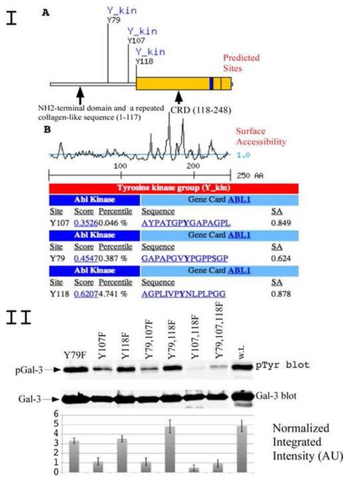
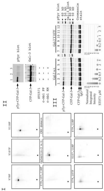
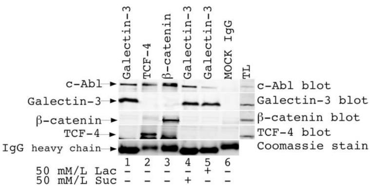
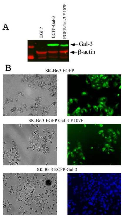
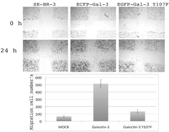
Galectin-3, a beta-galactoside-binding lectin, is found in cellular and extracellular location of the cell and has pleiotropic biological functions such as cell growth, cell adhesion and cell-cell interaction. It may exhibit anti- or pro-apoptotic activity depending on its localization and post-translational modifications. Two important post-translational modifications of galectin-3 have been reported: its cleavage and phosphorylation. Cleavage of galectin-3 was reported to be involved with angiogenic potential and apoptotic resistance. Phosphorylation of galectin-3 regulates its sugar-binding ability. In this report we have identified novel tyrosine phosphorylation sites in galectin-3 as well as the kinase responsible for its phosphorylation. Our results demonstrate that tyrosines at positions 79, 107 and 118 can be phosphorylated in vitro and in vivo by c-Abl kinase. Tyrosine 107 is the main target of c-Abl. Expression of galectin-3 Y107F mutant in galectin-3 null SK-Br-3 cells leads to morphological changes and increased motility compared to wild type galectin-3. Further investigation is needed to better understand the functional significance of the novel tyrosine phosphorylated sites of galectin-3.


Figures

Figure1
Figure1
I. Bioinformatic search predicting possible tyrosine phosphorylation sities of galectin-3 and the cognate protein kinase. (A) A diagram depicting newly identified galectin-3 phosphorylation sites and potential kinase. Indicated are NH2-terminal domain, a repeated collagen-like sequence and carbohydrate binding domain (CRD). (B) A diagram showing surface accessibility, scores and amino acids around predicted phosphorylation sites. II. c-Abl phosphorylation of galectin-3 in vitro. Recombinant GST-Galectin-3 mutants of predicted tyrosine sites and galectin-3 wild type were used as substrate for active c-Abl in in vitro assay. The reaction was stopped by adding sample buffer, resolved on 10% polyacrilomide gel, and immunoblotted using anti-pTyr antibody (top) or anti-galectin-3 antibody (bottom). The normalized integrated intensity was calculated as Band integrated intensity/Line normalization factor, anti-galectin-3 blot was used as reference channel.
Figure 2
Figure 2
I. Direct phosphorylation of galectin-3 by c-Abl in vitro. Approximately 1 μg of recombinant wild-type or mutated galectin-3 proteins used as substract for active c-Abl in in vitro assay. 100 μl kinase assay buffer containing 100 μM ATP and 25 μCi of [g-32P] ATP with recombinant galectin-3 and active c-Abl was incubated for 20 min at 30°C. After reaction was stopped with sample buffer, boiled samples were subjected to phosphopeptide map analysis as described in Materials and Methods. II. Phosphorylation of galectin-3 by c-Abl in vivo. Wild type galectin-3 protein was expressed in SK-Br-3 cell with constitutively active c-Abl PP (lane 1,2) and kinase dead c-Abl KM (lane3). Cells in lane 2 were treated with 10 μM of c-Abl inhibitor STI571. Galectin-3 was immunoprecipitated using anti-galectin-3 (TIB166) antibody and loaded on gel. The samples were resolved using 10% SDS-PAGE and immunoblotted with anti-Gal-3 (HL31) (top) and anti-phosphotyrosine antibodies (bottom). III. Dose-dependent inhibition of c-Abl activity by STI571. SK-Br-3 cells were co-transfected with c-Abl PP and wild type (lane 1-6) or Y107F (lane 7-12) galectin-3. STI571 was added to cell medium overnight. Cells were lysed and galectin-3 was immunoprecipitated using anti-Gal-3 (TIB166) antibody and loaded on gel. The samples were resolved using 10% SDS-PAGE gel and immunoblotted with anti-Gal-3 (HL31) (top) and anti-phosphotyrosine antibody (middle). To confirm equal loading of immunoprecipitated proteins 15% of IP mixture was run on 10% SDS-PAGE and visualized with Coomassie blue stain. The normalized integrated intensity was calculated as Band integrated intensity/Lane normalization factor, anti-galectin-3 blot was used as reference channel. The band having the highest integrated intensity is assigned a normalization factor of 1.0. This reference band is then used to calculate the normalization factor for all other selected lanes by dividing the band integrated intensity by the integrated intensity of the reference band. Then the normalization factor was used to calculate the normalized integrated intensity.
Figure 3
Figure 3
Coimmunoprecipitation of c-Abl with Gal-3. MDA-MB-435 cell lysates were immunoprecipitated using anti-galectin-3 (lanes 1,4,5), anti-TCF-4 (lane 2) and anti-β-catenin (lane 3) antibody and immunoblotted with anti-c-Abl, anti-TCF-4, anti-β-catenin and anti-galectin-3 polyclonal antibodies. Some cells were treated with sucrose and lactose (line 4,5 respectively). To confirm equal loading of immunoprecipitated proteins 15% of IP mixture was run on 10% SDS-PAGE and visualized with Coomassie blue stain.
Figure 4
Figure 4
Photomicrograph depicting the morphology of Sk-Br-3 stable cell lines expressing wild type and Y107F galectin-3. (A) Western blot analysis of Sk-Br-3 clones: transfected with EGFP (lane 1), ECFP-Gal-3 (lane 2) and EGFP-Gal-3 Y107F (lane 3). (B) Cellular shapes were visualized 2 days after splitting. Cells were grown under identical conditions. Left (phase contrast) and right (fluorescent light) panels represent same area on plate, x20.
Figure 5
Figure 5
Wound healing assay of SK-Br-3 cells were performed with stable cell lines constituvely expressing galectin-3 wild type and galectin-3 Y107F mutant. Cells were grown under identical conditions.

Similar articles

Cited by

References

    1. Barondes SH, Castronovo V, Cooper DN, Cummings RD, Drickamer K, Feizi T, Gitt MA, Hirabayashi J, Hughes C, Kasai K. Galectins: a family of animal beta-galactoside-binding lectins. Cell. 1994;76:597–598. - PubMed
    1. Barondes SH, Cooper DN, Gitt MA, Leffler H. Galectins. Structure and function of a large family of animal lectins. J. Biol. Chem. 1994;269:20807–20810. - PubMed
    1. Califice S, Castronovo V, Van Den Brule F. Galectin-3 and cancer. Int. J. Oncol. 2004;25:983–992. - PubMed
    1. Dumic J, Dabelic S, Flogel M. Galectin-3: an open-ended story. Biochim. Biophys. Acta. 2006;1760:616–635. - PubMed
    1. Huflejt ME, Turck CW, Lindstedt R, Barondes SH, Leffler H. L-29, a soluble lactose-binding lectin, is phosphorylated on serine 6 and serine 12 in vivo and by casein kinase I. J. Biol. Chem. 1993;268:26712–26718. - PubMed

Publication types

MeSH terms