

Focused ultrasound and microbubbles for enhanced extravasation
- PMID: 20600402
- PMCID: PMC3085646
- DOI: 10.1016/j.jconrel.2010.06.012
Focused ultrasound and microbubbles for enhanced extravasation
Abstract
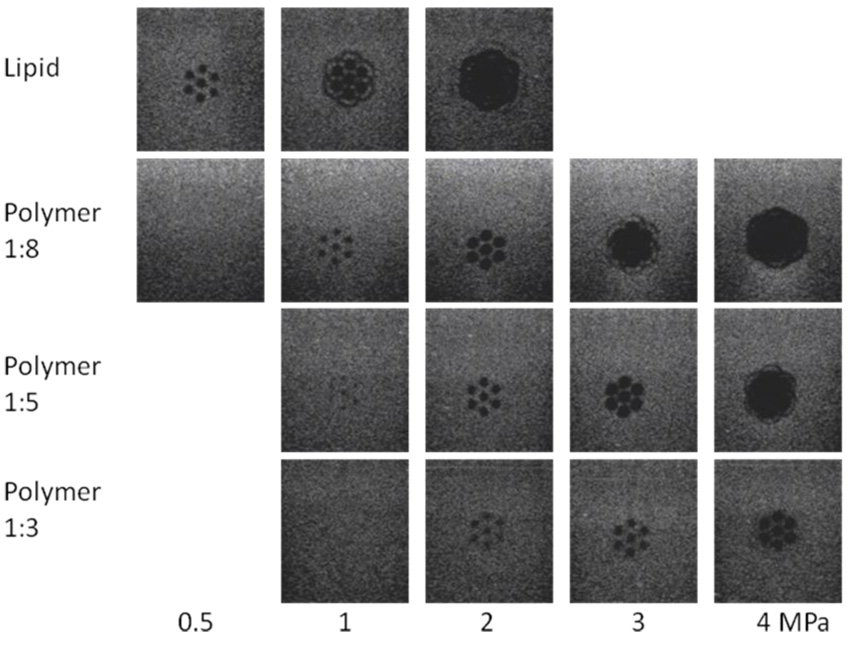
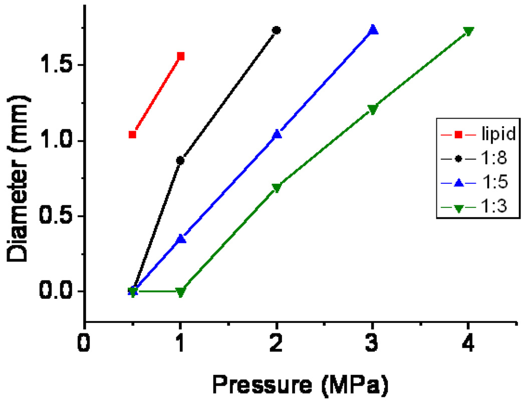
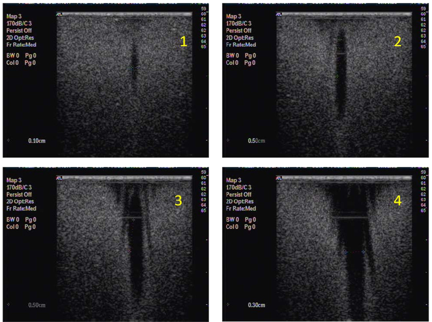
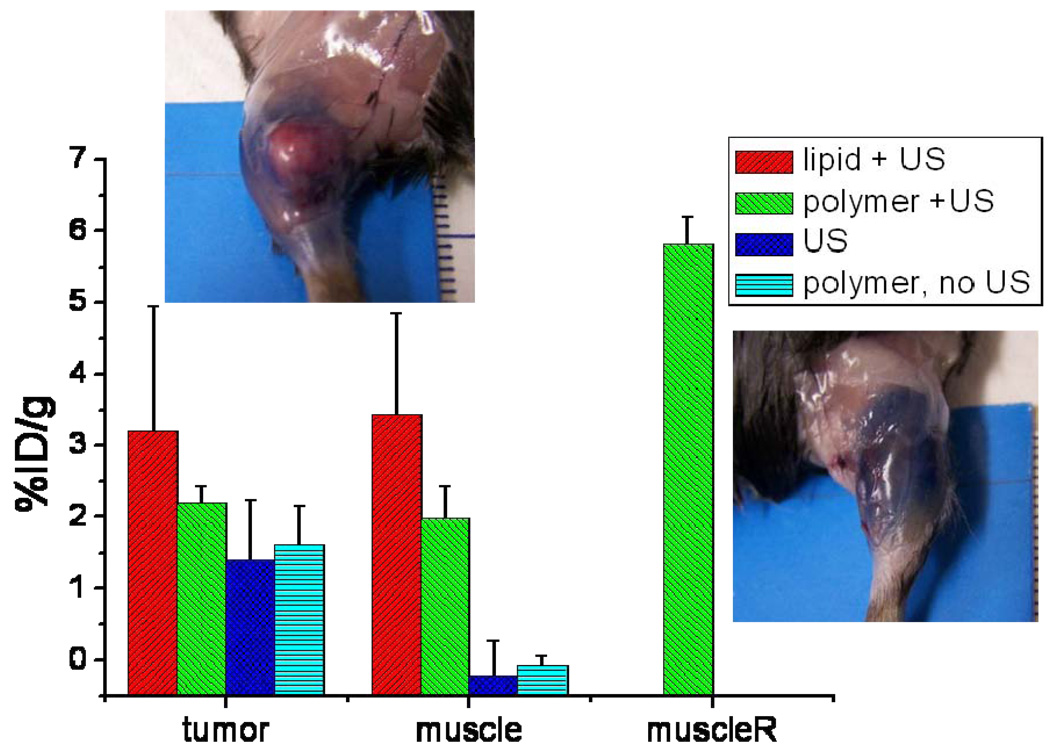
The permeability of blood vessels for albumin can be altered by using ultrasound and polymer or lipid-shelled microbubbles. The region in which the microbubbles were destroyed with focused ultrasound was quantified in gel phantoms as a function of pressure, number of cycles and type of microbubble. At 2MPa the destruction took place in a fairly wide area for a lipid-shelled agent, while for polymer-shelled agents at this setting, distinct destruction spots with a radius of only 1mm were obtained. When microbubbles with a thicker shell were used, the pressure above which the bubbles were destroyed shifts to higher values. In vivo both lipid and polymer microbubbles increased the extravasation of the albumin binding dye Evans Blue, especially in muscle leading to about 6-8% of the injected dose to extravasate per gram muscle tissue 30 min after start of the treatment, while no Evans Blue could be detected in muscle in the absence of microbubbles. Variation in the time between ultrasound treatment and Evans Blue injection, demonstrated that the time window for promoting extravasation is at least an hour at the settings used. In MC38 tumors, extravasation already occurred without ultrasound and only a trend towards enhancement with about a factor of 2 could be established with a maximum percentage injected dose per gram of 3%. Ultrasound mediated microbubble destruction especially enhances the extravasation in the highly vascularized outer part of the MC38 tumor and adjacent muscle and would, therefore, be most useful for release of, for instance, anti-angiogenic drugs.
Copyright © 2010 Elsevier B.V. All rights reserved.
Figures
References
-
- Nixdorff U, Schmidt A, Morant T, Stilianakis TN, Voigt JU, Flachskampf FA, Daniel WG, Garlichs CD. Dose-dependent disintegration of human endothelial monolayers by contrast echocardiography. Life Sciences. 2005;77:1493–1501. - PubMed
-
- Stieger SM, Caskey CF, Adamson RH, Qin SP, Curry FRE, Wisner ER, Ferrara KW. Enhancement of vascular permeability with low-frequency contrast-enhanced ultrasound in the chorioallantoic membrane model. Radiology. 2007;243:112–121. - PubMed
-
- Song J, Chappell JC, Qi M, VanGieson EJ, Kaul S, Price RJ. Influence of injection site, microvascular pressure and ultrasound variables on microbubble-mediated delivery of microspheres to muscle. Journal of the American College of Cardiology. 2002;39:726–731. - PubMed
-
- Price RJ, Skyba DM, Kaul S, Skalak TC. Delivery of colloidal particles and red blood cells to tissue through microvessel ruptures created by targeted microbubble destruction with ultrasound. Circulation. 1998;98:1264–1267. - PubMed
-
- Geis NA, Mayer CR, Kroll RD, Hardt SE, Katus HA, Bekeredjian R. Spatial distribution of ultrasound targeted microbubble destruction increases cardiac transgene expression but not capillary permeability. Ultrasound Med Biol. 2009;35:1119–1126. - PubMed
Publication types
MeSH terms
Substances
Grants and funding
LinkOut - more resources
Full Text Sources
Other Literature Sources
