Review
. 2010 Nov-Dec;28(6):770-81.
doi: 10.1016/j.biotechadv.2010.06.004. Epub 2010 Jul 1.

Advances in developing HIV-1 viral load assays for resource-limited settings

Affiliations
Review

Advances in developing HIV-1 viral load assays for resource-limited settings

ShuQi Wang et al. Biotechnol Adv. 2010 Nov-Dec.

Abstract

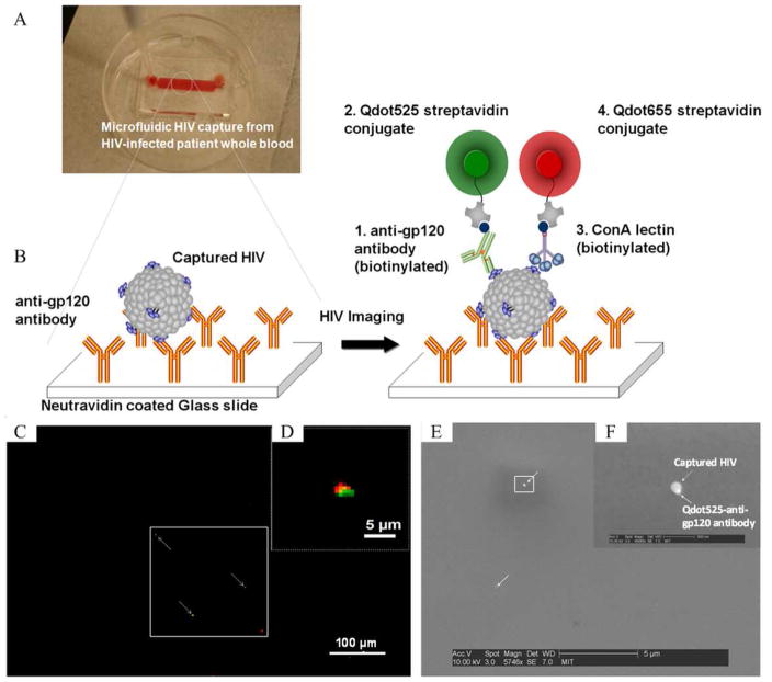
Commercial HIV-1 RNA viral load assays have been routinely used in developed countries to monitor antiretroviral treatment (ART). However, these assays require expensive equipment and reagents, well-trained operators, and established laboratory infrastructure. These requirements restrict their use in resource-limited settings where people are most afflicted with the HIV-1 epidemic. Inexpensive alternatives such as the Ultrasensitive p24 assay, the reverse transcriptase (RT) assay and in-house reverse transcription quantitative polymerase chain reaction (RT-qPCR) have been developed. However, they are still time-consuming, technologically complex and inappropriate for decentralized laboratories as point-of-care (POC) tests. Recent advances in microfluidics and nanotechnology offer new strategies to develop low-cost, rapid, robust and simple HIV-1 viral load monitoring systems. We review state-of-the-art technologies used for HIV-1 viral load monitoring in both developed and developing settings. Emerging approaches based on microfluidics and nanotechnology, which have potential to be integrated into POC HIV-1 viral load assays, are also discussed.


Figures

Figure 1
Figure 1. Global distribution of HIV-1 infection in 2007 (adapated from UNAIDS, 2008)
By the end of 2007, it is estimated that 33 million people were infected with HIV-1. Sub-Saharan Africa accounts for 67 % of infections, with prevalence in adults up to 28 %. Main subtypes are shown as previously reported (Taylor et al., 2008).
Figure 2
Figure 2. Diagnostic markers during the natural course of HIV-1 infection (adapted from Chang et al., 2006)
The natural course of HIV-1 infection can be divided into three stages: seroconversion, asymptomatic and symptomatic. HIV-1 RNA (red line) peaks during seroconversion, then decreases and remains at a low level and shows no clinical symptoms. HIV-1 p24 antigen (blue triangles) demonstrates the same trend as HIV-1 RNA. HIV-1 specific antibody (green circles) is produced during seroconversion and reaches a plateau at the asymptomatic stage. The number of CD4+ cells (black squares) drops rapidly and rebounds during seroconversion and it gradually decrease as AIDS develops.
Figure 3
Figure 3. Schematic illustration of the TaqMan technology for quantification (reproduced from Liegler and Grant, 2006)
(A) Principle for PCR amplification and the TaqMan probe. In each cycle of PCR, a new DNA fragment is amplified by DNA polymerase with the aid of forward and reverse primers. During amplification, a TaqMan probe binding to the DNA template is digested by DNA polymerase, releasing fluorescence. (B) Amplification plots of quantification standards and an unknown sample. Six external standards are amplified in parallel with an unknown sample. The increase of fluorescence from each reaction is recorded during the amplification. (C) Quantification of the unknown sample by comparing with the standard curve. The CT values (where fluorescence of each reaction exceeds the background) of quantification standards are plotted against HIV-1 viral load. The viral load of the unknown sample is indicated by the green check.
Figure 4
Figure 4. Schematic illustration of the BCA technology for DNA detection (reproduced from Nam et al., 2003)
(A) Functionalization of gold nanoparticles (NPs) and magnetic microparticles (MMPs). NPs (in pink) are dual-labeled with polyclonal antibody against prostate-specific antigen (PSA, in blue) and many copies of barcode DNA (in green). MMPs (in grey) are heavily conjugated to PSA monoclonal antibody (in red). (B) Barcode DNA mediated protein separation and scanometric detection with/without nucleic acid amplification by PCR.
Figure 5
Figure 5. A microfluidics-based viral load device to capture and image HIV-1 particles (adapted from Kim et al., 2009)
(A) A top-down image of a microfluidic device containing whole blood. (B) Schematic illustration of the HIV-1 capturing/imaging strategy. On a glass slide, anti-gp120 antibody is immobilized. HIV-1 particles, which have gp120 antigen protruding on the surface, are captured via specific antigen-antibody interaction. The captured particles are then co-recognized by biotinylated anti-gp120 and ConA lectin, which are conjugated to quantum dots 525 (solid green circle) and 655 (solid red circle), respectively. (C–D) Under a fluorescence microscope, HIV-1 particles are identified by co-located green and red light, and quantified using an imaging system. (E–F) SEM images of the captured HIV particle with Qdot525/anti-gp120 antibody.

References

    1. Abbott. Abbott RealTime HIV-1 (package insert) 2007.
    1. Alyassin MA, Moon S, Keles HO, Manzur F, Lin RL, Haeggstrom E, et al. Rapid automated cell quantification on HIV microfluidic devices. Lab Chip. 2009;9:3364–9. - PMC - PubMed
    1. Bhattacharyya A, Klapperich CM. Design and testing of a disposable microfluidic chemiluminescent immunoassay for disease biomarkers in human serum samples. Biomed Microdevices. 2007;9:245–51. - PubMed
    1. Bonard D, Rouet F, Toni TA, Minga A, Huet C, Ekouevi DK, et al. Field evaluation of an improved assay using a heat-dissociated p24 antigen for adults mainly infected with HIV-1 CRF02_AG strains in Cote d’Ivoire, West Africa. J Acquir Immune Defic Syndr. 2003;34:267–73. - PubMed
    1. Boni J, Opravil M, Tomasik Z, Rothen M, Bisset L, Grob PJ, et al. Simple monitoring of antiretroviral therapy with a signal-amplification-boosted HIV-1 p24 antigen assay with heat-denatured plasma. Aids. 1997;11:F47–52. - PubMed

Publication types

LinkOut - more resources