

Macroautophagy is defective in mucolipin-1-deficient mouse neurons
- PMID: 20600908
- PMCID: PMC4392647
- DOI: 10.1016/j.nbd.2010.06.010
Macroautophagy is defective in mucolipin-1-deficient mouse neurons
Abstract
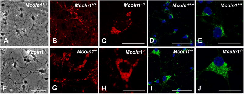
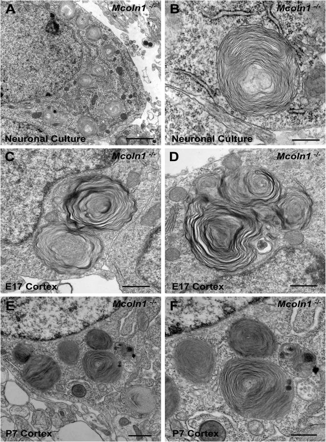
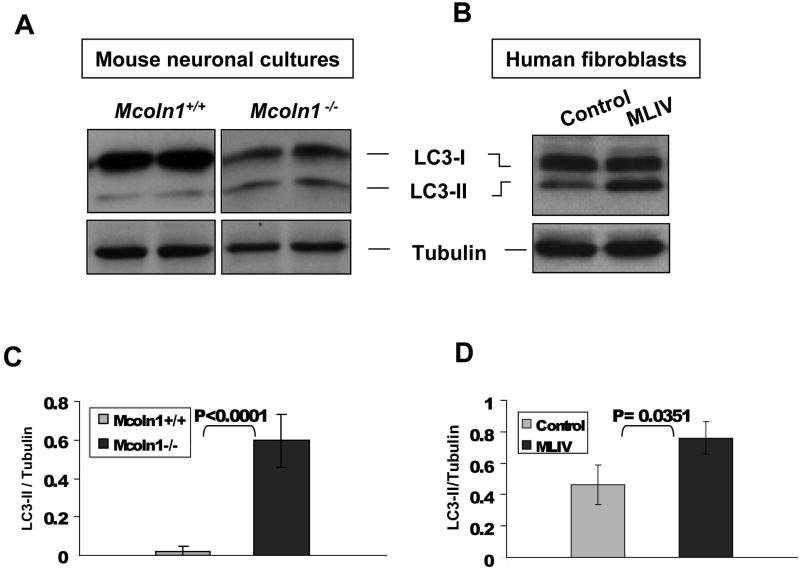
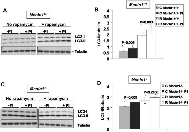
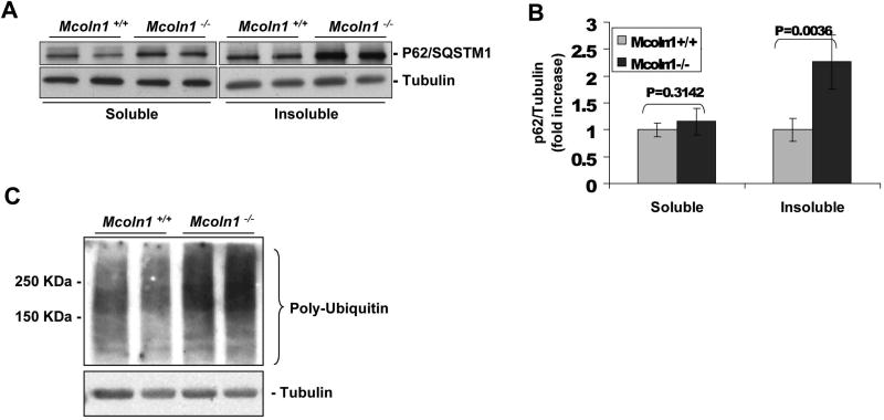
Mucolipidosis type IV is a neurodegenerative lysosomal disease clinically characterized by psychomotor retardation, visual impairment, and achlorhydria. In this study we report the development of a neuronal cell model generated from cerebrum of Mcoln1(-/-) embryos. Prior functional characterization of MLIV cells has been limited to fibroblast cultures gleaned from patients. The current availability of the mucolipin-1 knockout mouse model Mcoln1(-/-) allows the study of mucolipin-1-defective neurons, which is important since the disease is characterized by severe neurological impairment. Electron microscopy studies reveal significant membranous intracytoplasmic storage bodies, which correlate with the storage morphology observed in cerebral cortex of Mcoln1(-/-) P7 pups and E17 embryos. The Mcoln1(-/-) neuronal cultures show an increase in size of LysoTracker and Lamp1 positive vesicles. Using this neuronal model system, we show that macroautophagy is defective in mucolipin-1-deficient neurons and that LC3-II levels are significantly elevated. Treatment with rapamycin plus protease inhibitors did not increase levels of LC3-II in Mcoln1(-/-) neuronal cultures, indicating that the lack of mucolipin-1 affects LC3-II clearance. P62/SQSTM1 and ubiquitin levels were also increased in Mcoln1(-/-) neuronal cultures, suggesting an accumulation of protein aggregates and a defect in macroautophagy which could help explain the neurodegeneration observed in MLIV. This study describes, for the first time, a defect in macroautophagy in mucolipin-1-deficient neurons, which corroborates recent findings in MLIV fibroblasts and provides new insight into the neuronal pathogenesis of this disease.
Copyright © 2010 Elsevier Inc. All rights reserved.
Figures
Similar articles
-
Neuropathology of the Mcoln1(-/-) knockout mouse model of mucolipidosis type IV.J Neuropathol Exp Neurol. 2009 Feb;68(2):125-35. doi: 10.1097/NEN.0b013e3181942cf0. J Neuropathol Exp Neurol. 2009. PMID: 19151629 Free PMC article.
-
Chaperone-mediated autophagy is defective in mucolipidosis type IV.J Cell Physiol. 2009 May;219(2):344-53. doi: 10.1002/jcp.21676. J Cell Physiol. 2009. PMID: 19117012
-
Autophagic dysfunction in mucolipidosis type IV patients.Hum Mol Genet. 2008 Sep 1;17(17):2723-37. doi: 10.1093/hmg/ddn174. Epub 2008 Jun 11. Hum Mol Genet. 2008. PMID: 18550655 Free PMC article.
-
The molecular basis of mucolipidosis type IV.Curr Mol Med. 2002 Aug;2(5):445-50. doi: 10.2174/1566524023362276. Curr Mol Med. 2002. PMID: 12125810 Review.
-
Mucolipin 1: endocytosis and cation channel--a review.Pflugers Arch. 2005 Oct;451(1):313-7. doi: 10.1007/s00424-004-1361-7. Epub 2004 Nov 27. Pflugers Arch. 2005. PMID: 15570434 Review.
Cited by
-
The mucolipidosis IV Ca2+ channel TRPML1 (MCOLN1) is regulated by the TOR kinase.Biochem J. 2015 Sep 15;470(3):331-42. doi: 10.1042/BJ20150219. Epub 2015 Jul 20. Biochem J. 2015. PMID: 26195823 Free PMC article.
-
Lysosomal physiology.Annu Rev Physiol. 2015;77:57-80. doi: 10.1146/annurev-physiol-021014-071649. Annu Rev Physiol. 2015. PMID: 25668017 Free PMC article. Review.
-
The first genetically confirmed Japanese patient with mucolipidosis type IV.Clin Case Rep. 2016 Apr 13;4(5):509-12. doi: 10.1002/ccr3.540. eCollection 2016 May. Clin Case Rep. 2016. PMID: 27190617 Free PMC article.
-
The role of autophagy in neurodegenerative disease.Nat Med. 2013 Aug;19(8):983-97. doi: 10.1038/nm.3232. Epub 2013 Aug 6. Nat Med. 2013. PMID: 23921753 Review.
-
Lysosomal TRPML1 regulates mitochondrial function in hepatocellular carcinoma cells.J Cell Sci. 2022 Mar 15;135(6):jcs259455. doi: 10.1242/jcs.259455. Epub 2022 Mar 21. J Cell Sci. 2022. PMID: 35274126 Free PMC article.
References
-
- Bargal R, Avidan N, Ben-Asher E, Olender Z, Zeigler M, Frumkin A, et al. Identification of the gene causing mucolipidosis type IV. Nat Genet. 2000;26:118–23. - PubMed
-
- Berman ER LN, Shapira E, Merin S, Levij IS. Congenital corneal clouding with abnormal systemic storage bodies: a new variant of mucolipidosis. J Pediatr. 1974;84:519–26. - PubMed
-
- Brewer GJ, Torricelli JR, Evege EK, Price PJ. Optimized survival of hippocampal neurons in B27-supplemented Neurobasal, a new serum-free medium combination. J Neurosci Res. 1993;35:567–76. - PubMed
Publication types
MeSH terms
Substances
Grants and funding
LinkOut - more resources
Full Text Sources
Molecular Biology Databases
Miscellaneous
