. 2010 Oct;45(2):163-72.
doi: 10.1016/j.mcn.2010.06.007. Epub 2010 Jun 21.

Conantokins inhibit NMDAR-dependent calcium influx in developing rat hippocampal neurons in primary culture with resulting effects on CREB phosphorylation

Affiliations

Conantokins inhibit NMDAR-dependent calcium influx in developing rat hippocampal neurons in primary culture with resulting effects on CREB phosphorylation

Luoxiu Huang et al. Mol Cell Neurosci. 2010 Oct.

Abstract

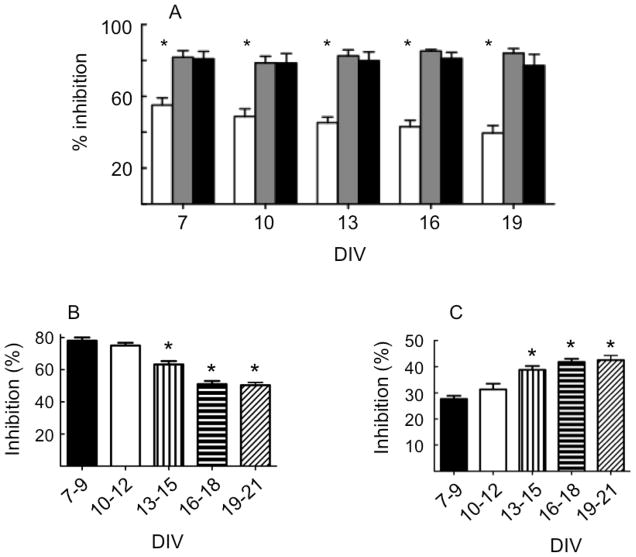
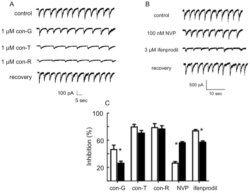
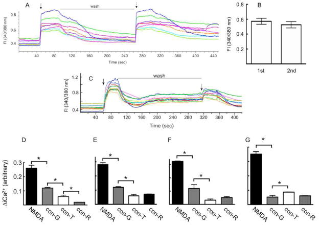
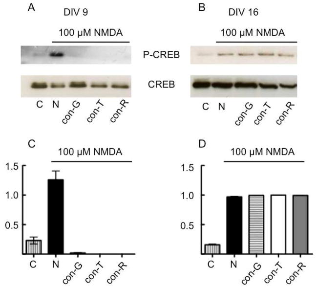
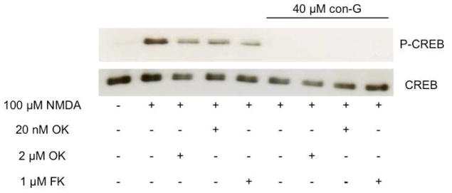
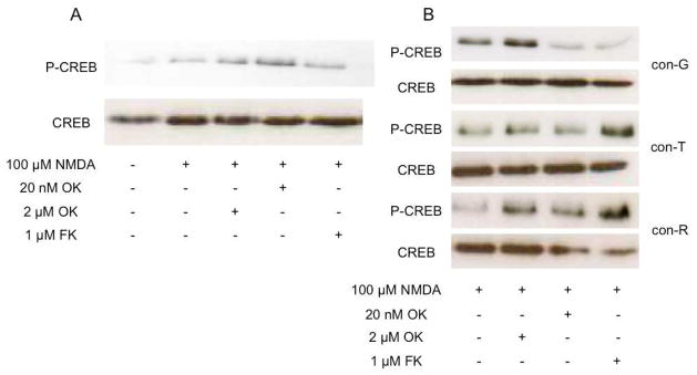
The effects of conantokin (con)-G, con-R[1-17], and con-T on ion flow through N-methyl-D-aspartate receptor (NMDAR) ion channels were determined in cultured primary rat hippocampal neurons. The potency of con-G diminished, whereas inhibition by con-R[1-17] and con-T did not change, as the neurons matured. Con-G, con-R[1-17], and con-T effectively diminished NMDA-induced Ca(2+) influx into the cells. A similar age-dependent decrease in con-G-mediated inhibition of the amplitude of spontaneous excitatory postsynaptic currents (sEPSCs) was observed, compared to con-R[1-17] and con-T. The effects of the conantokins on NMDA-induced cAMP response element-binding protein (CREB) phosphorylation in immature (DIV 9) and mature (DIV 16) neurons showed that, at DIV 9, con-G, con-R[1-17], and con-T inhibited NMDA-mediated P-CREB levels, whereas in DIV 16 neurons the conantokins did not inhibit overall levels of NMDA-induced P-CREB. In contrast, P-CREB levels were enhanced through inhibition of the protein phosphatases, PP1 and PP2B (calcineurin). This ability of conantokins to sustain CREB phosphorylation can thus enhance neuronal survival and plasticity.


Conflict of interest statement

Conflict of interest statement

The authors declare no conflict of interest.

Figures

Fig. 1
Fig. 1
The inhibition by 20 μM con-G (white bars), 20 μM con-R[1-17] (black bars), and 20 μM con-T (grey bars), of NMDA-evoked currents as a function of developmental age of the neurons (*p<0.05 between con-G and either con-R[1-17] or con-T; n = 6-9 at each time). B, C. Age-dependency of ifenprodil and NVP-AAM077-inhibition of NMDA evoked whole cell currents in developing neurons. The changes in the relative inhibition by 3 μM ifenprodil (B) and 100 nM NVP (C) as a function of neuronal age in culture (*p<0.05 compared to DIV 7-9, n = 7-11 at each age).
Fig. 2
Fig. 2
Inhibition of the sEPSCs of developing neurons by conantokins. An example of NMDAR-derived sEPSCs recorded from hippocampal neurons at DIV 14. The control buffer was 10 μM glycine, 20 μM bicuculline, 1 μM strychnine, and 20 μM CNQX. A. Conantokins inhibited the peak amplitudes of the sEPSCs, and this inhibition was reversible after wash-out of these inhibitors. B. Similar data are shown for inhibition of NMDA-elicited sEPSCs by 100 nM NVP-AAM077 (NVP) and 3 μM ifenprodil. C. Quantitation of the inhibition of the peak amplitudes of the sEPSCs of DIV 14 (white bars) and DIV 19 (black bars) neurons by various inhibitors (*p<0.05 for pairwise comparisons, n =7-11).
Fig. 3
Fig. 3
Single cell iCa2+ measurements upon NMDA stimulation in the absence or presence of conantokins. (A). An example of the imaging of iCa2+ transients after stimulation (solid arrow) of DIV 13 neurons with 50 αM NMDA/10 αM glycine as monitored by the fluorescence (Fl) ratio at 340/380 nm. After wash-out (wash) of 50 αM NMDA/10 αM glycine, the neurons were reexposed to the same stimulation (dashed arrow) to assess the viability of the neurons to a second application of coagonists. (B). The maximum Fl 340/380 ratio as a result of the 1st and 2nd stimulation by 50 αM NMDA/10 αM glycine, showing that the neurons maintained the same level of viability. (C). Inhibition of the iCa2+ transients of DIV 13 neurons as a result of application of con-G. After washout of 50 αM NMDA/10 αM glycine from the first stimulation (solid arrow), the neurons were restimulated with 50 αM NMDA/10 αM glycine/40 αM con-G. A decrease in the maximum Fl (340/380) is seen, quantitatively demonstrating the effectiveness of con-G. (D-G). The effect of con-G, con-R[1-17] (con-R), and con-T on the increase in [iCa2+] at DIV 7 (D), DIV 13 (E), DIV 16 (F), and DIV 19 (G). The data are expressed as the mean ± S.E.M. of 3 independent experiments with 10–13 neurons per experiment. Statistical analysis * P < 0.05 compares iCa2+ changes between NMDA alone and NMDA/con-G, NMDA alone and NMDA/con-R[1-17], and NMDA alone and NMDA/con-T.
Fig. 4
Fig. 4
Inhibition of CREB phosphorylation by con-G, con-R[1-17] (con-R), and con-T. Cultured hippocampal neurons at DIV 9 (A) and DIV 16 (B) were stimulated with 100 αM NMDA or 100 αM NMDA/40 αM conantokins for 20 min. Total cell lysates were then obtained and immunoblotted for P-CREB, CREB, and the loading control, tubulin. C, D. Densitometric analysis is provided of the blots representing the ratio of P-CREB/CREB for treatments performed on neurons at (A) DIV 9 and (B) DIV 16.
Fig. 5
Fig. 5
Effect of protein phosphatase inhibitors on levels of P-CREB. Cultured hippocampal neurons at DIV 9 were treated with 100 αM NMDA in the presence or absence of 40 αM con-G for 5 min and then treated with the indicated phosphatase inhibitors for 20 min. Since nearly identical data were obtained with con-R[1-17], con-T, and MK-801 for DIV 9 neurons, only the immunoblot with con-G is shown as an example. Total cell lysates were immunoblotted for P-CREB and CREB.
Fig. 6
Fig. 6
Effects of NMDAR antagonists on levels of P-CREB. Neurons at DIV 16 were treated with 100 αM NMDA (A) or 100 αM NMDA/conantokins (B) for 5 min, and then treated with the indicated phosphatase inhibitors for 20 min. Total cell lysates were immunoblotted for P-CREB and CREB.
Fig. 7
Fig. 7
Subcellular localization of NR2B in cultured hippocampal neurons after treatment with NMDA and NMDA/con-G at DIV 9 and DIV 16. The neurons were fixed and immunolabeled for NR2B (red) and CaMKII (green). (A, B). Control neurons at DIV 9 (A) and DIV 16 (B) pretreated with TTX, CNQX, and nifedipine. (C, D). DIV 9 (C) and DIV 16 (D) neurons treated with 100 αM NMDA for 20 min. (E, F). DIV 9 (E) and DIV 16 (F) neurons treated with 100 αM NMDA/40 αM con-G for 20 min. Panels (G-L) show only the NR2B staining (red) of images A-G. (G, H). Control neurons at DIV 9 (G) and DIV 16 (H). (I, J). DIV 9 (I) and DIV 16 (J) neurons treated with 100 αM NMDA for 20 min. (K, L). DIV 9 (K) and DIV 16 (L) neurons treated with 100 αM NMDA/40 αM con-G for 20 min. The solid white arrows show examples of NR2B staining in the dendrites and broken white arrows indicate examples of staining of NR2B in cell bodies. The intense red staining around the nucleus of the cells is an antibody artifact observed in many studies of this type.

References

    1. Alex AB, Baucum AJ, Wilcox KS. Effect of Conantokin G on NMDA receptor-mediated spontaneous EPSCs in cultured cortical neurons. J Neurophysiol. 2006;96:1084–1092. - PubMed
    1. Auberson YP, Allgeier H, Bischoff S, Lingenhoehl K, Moretti R, Schmutz M. 5-phosphonomethylquinoxalinediones as competitive NMDA receptor antagonists with a preference for the human 1A/2A, rather than 1A/2B receptor composition. Bioorg Med Chem Lett. 2002;12:1099–1102. - PubMed
    1. Barria A, Malinow R. NMDA receptor subunit composition controls synaptic plasticity by regulating binding to CaMKII. Neuron. 2005;48:289–301. - PubMed
    1. Barton ME, White HS, Wilcox KS. The effect of CGX-1007 and CI-1041, novel NMDA receptor antagonists, on NMDA receptor-mediated EPSCs. Epilepsy Res. 2004;59:13–24. - PubMed
    1. Bialojan C, Takai A. Inhibitory effect of a marine-sponge toxin, okadaic acid, on protein phosphatases. Specificity and kinetics. Biochem J. 1988;256:283–290. - PMC - PubMed

MeSH terms