. 2010 Aug;7(8):637-42.
doi: 10.1038/nmeth.1476. Epub 2010 Jul 4.

Fast, high-contrast imaging of animal development with scanned light sheet-based structured-illumination microscopy

Affiliations

Fast, high-contrast imaging of animal development with scanned light sheet-based structured-illumination microscopy

Philipp J Keller et al. Nat Methods. 2010 Aug.

Abstract

Recording light-microscopy images of large, nontransparent specimens, such as developing multicellular organisms, is complicated by decreased contrast resulting from light scattering. Early zebrafish development can be captured by standard light-sheet microscopy, but new imaging strategies are required to obtain high-quality data of late development or of less transparent organisms. We combined digital scanned laser light-sheet fluorescence microscopy with incoherent structured-illumination microscopy (DSLM-SI) and created structured-illumination patterns with continuously adjustable frequencies. Our method discriminates the specimen-related scattered background from signal fluorescence, thereby removing out-of-focus light and optimizing the contrast of in-focus structures. DSLM-SI provides rapid control of the illumination pattern, exceptional imaging quality and high imaging speeds. We performed long-term imaging of zebrafish development for 58 h and fast multiple-view imaging of early Drosophila melanogaster development. We reconstructed cell positions over time from the Drosophila DSLM-SI data and created a fly digital embryo.


Figures

Figure 1
Figure 1. Light sheet-based structured illumination with digitally adjustable frequency
(a) Side-view of the central components of a Digital Scanned Laser Light Sheet Fluorescence Microscope (DSLM). The DSLM illumination lens illuminates a thin volume by rapidly scanning a µm-sized laser beam through the specimen. Fluorescence is detected at a right angle to the illuminated plane by the detection lens. The intensity of the laser beam is modulated in synchrony with the scanning process. (b) Cross-sections of DSLM/DSLM-SI illumination profiles, recorded at 10× magnification. A uniform intensity distribution is used in the standard DSLM light sheet illumination mode (LS, first profile). In the DSLM structured illumination mode (SI), the spatial laser light intensity distribution is modulated in a sinusoidal fashion. The patterns shown here as reflections of a mirror oriented at 45 ° to the incident laser beam range from four sine periods (SI-4) to 180 sine periods (SI-180) across the field-of-view. The width of the image corresponds to a field size of 1.5 mm. Center-to-center distances of the maxima in the patterns range between 412.5 µm (SI frequency s = 4, SI-4) and 9.2 µm (SI frequency s = 180, SI-180).
Figure 2
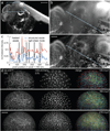
Figure 2. Enhancing image contrast with DSLM structured illumination
(a) Maximum-intensity projection of a DSLM image stack of a 3.5-day old Medaka fish embryo with Sytox Green nuclear staining, recorded with standard light sheet illumination (10× magnification). Stack dimensions: 435 images, recorded at a z-spacing of 3 µm, covering a total volume of 1,516 × 1,516 × 1,305 µm3. (b) Close-ups of the regions indicated in (a), recorded in standard light sheet mode (top) and in structured illumination mode (SI-32, bottom), respectively. (c) Intensity plot along the lines indicated in (b). In both plots, the raw intensity values are normalized by the same factor (global maximum of both images). Arrows and shading indicate structures in the structured illumination image that are not visible in the light sheet image. (d) Fast multi-channel imaging of early zebrafish embryogenesis with DSLM-SI: Maximum-intensity projections of a DSLM time-lapse recording of a membrane- and nuclei-labeled zebrafish embryo. The embryo was injected with ras-eGFP mRNA and H2A-mCherry mRNA at the 1-cell stage. Membranes were imaged using structured illumination (SI-25), nuclei using standard light sheet illumination (LS). In total, 125,280 images were recorded at 261 time points from 2 to 8.5 h.p.f.. Images were deconvolved with ten iterations of the Lucy-Richardson algorithm. Scale-bars = 200 µm (a), 50 µm (b), 100 µm (d). Carl Zeiss C-Apochromat 10×/0.45 W. Recording speed: 6 DSLM images per second, 2 SI-reconstructed images per second.
Figure 3
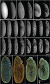
Figure 3. Multiple-view imaging of Drosophila embryogenesis with DSLM-SI
(a) Comparison of maximum-intensity projections of DSLM image stacks of a nuclei-labeled Drosophila embryo, using standard light sheet illumination (LS, left) and structured illumination (SI-20, right), respectively. The image pairs show the same volume at the same time point. (b) Maximum-intensity projections of a DSLM-SI multiple-view time-lapse recording of a nuclei-labeled Drosophila embryo. In total, 137,520 images were recorded at 191 time points from 2 to 11.5 h.p.f.. Images were deconvolved with the Lucy-Richardson algorithm (5 iterations). (c) Reconstructing Drosophila embryogenesis from DSLM-SI data: The panel shows lateral snapshots of the Fly Digital Embryo. Nuclei were automatically detected in the four views of the developing Drosophila embryo. The resulting point clouds were fused. Color-code: directed regional nuclei movement speeds over 10-min-periods. Scale-bars = 100 µm. Carl Zeiss Plan-Apochromat 20×/1.0 W. Recording speed: 6 DSLM images per second, 2 SI-reconstructed images per second.
Figure 4
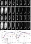
Figure 4. Spatiotemporal image contrast optimization by DSLM-SI frequency chirping
(a) DSLM images of a nuclei-labeled stage 5 Drosophila embryo (before internal structures have developed) at two different depths inside the specimen, using standard light sheet illumination and structured illumination patterns with different frequencies. The dashed line separates the region containing the background and out-of-focus signal that has been removed by DSLM-SI (left) from the in-focus structures where the contrast has been enhanced (right). (b) Planes as in (a), but at a later time point (stage 8). (c) Quantification of image contrast as a function of SI frequency and imaging depth (z) inside the specimen, for the time point shown in (a). (d) Same as in (c), but for the later time point shown in (b). Scale-bar = 100 µm. Carl Zeiss Plan-Apochromat 20×/1.0 W. Recording speed: 5 DSLM images per second, 1.7 SI-reconstructed images per second.

Similar articles

Cited by

References

    1. Bao Z, et al. Automated cell lineage tracing in Caenorhabditis elegans. Proceedings of the National Academy of Sciences of the United States of America. 2006;103:2707–2712. - PMC - PubMed
    1. Megason SG, Fraser SE. Imaging in systems biology. Cell. 2007;130:784–795. - PubMed
    1. Fowlkes CC, et al. A quantitative spatiotemporal atlas of gene expression in the Drosophila blastoderm. Cell. 2008;133:364–374. - PubMed
    1. McMahon A, Supatto W, Fraser SE, Stathopoulos A. Dynamic analyses of Drosophila gastrulation provide insights into collective cell migration. Science. 2008;322:1546–1550. - PMC - PubMed
    1. Murray JI, et al. Automated analysis of embryonic gene expression with cellular resolution in C. elegans. Nature Methods. 2008;5:703–709. - PMC - PubMed

Publication types

MeSH terms

LinkOut - more resources