. 2010 Aug;42(8):668-75.
doi: 10.1038/ng.613. Epub 2010 Jul 4.

Androgen-induced TOP2B-mediated double-strand breaks and prostate cancer gene rearrangements

Affiliations

Androgen-induced TOP2B-mediated double-strand breaks and prostate cancer gene rearrangements

Michael C Haffner et al. Nat Genet. 2010 Aug.

Abstract

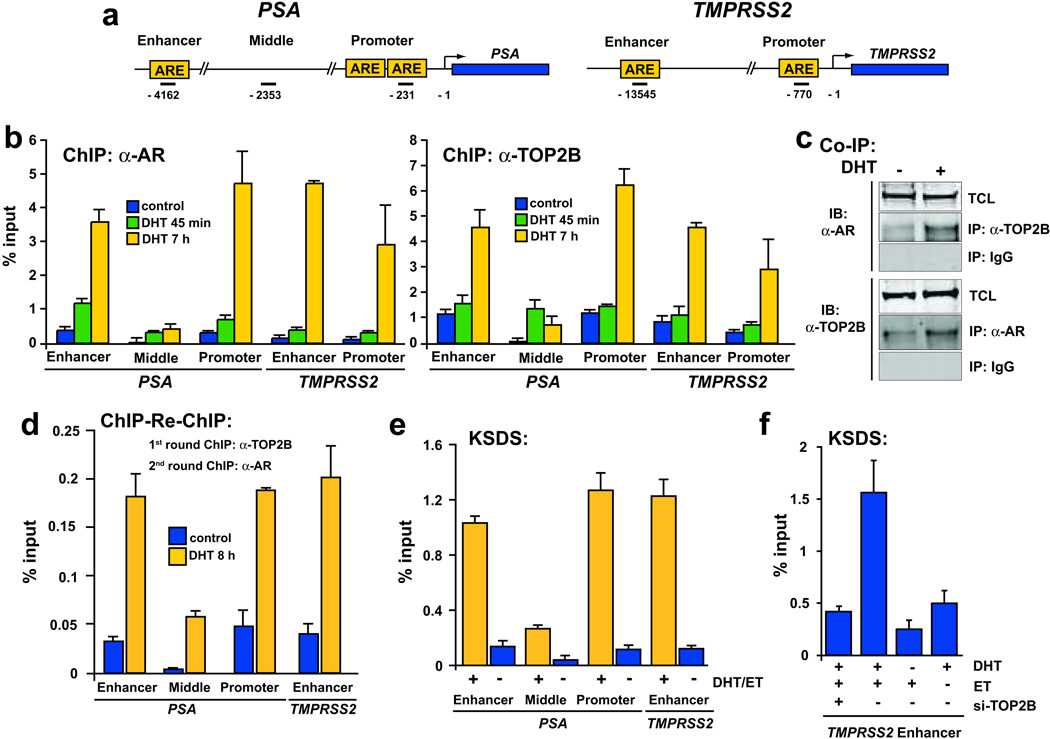
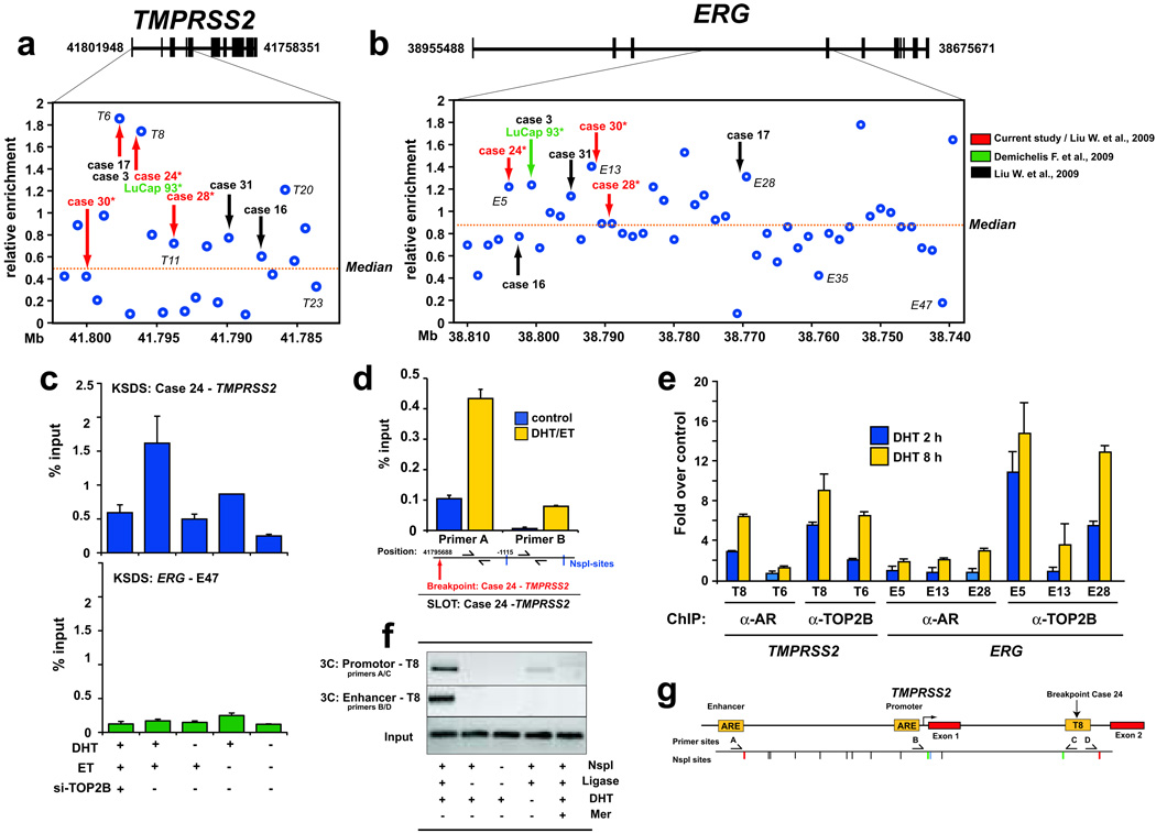
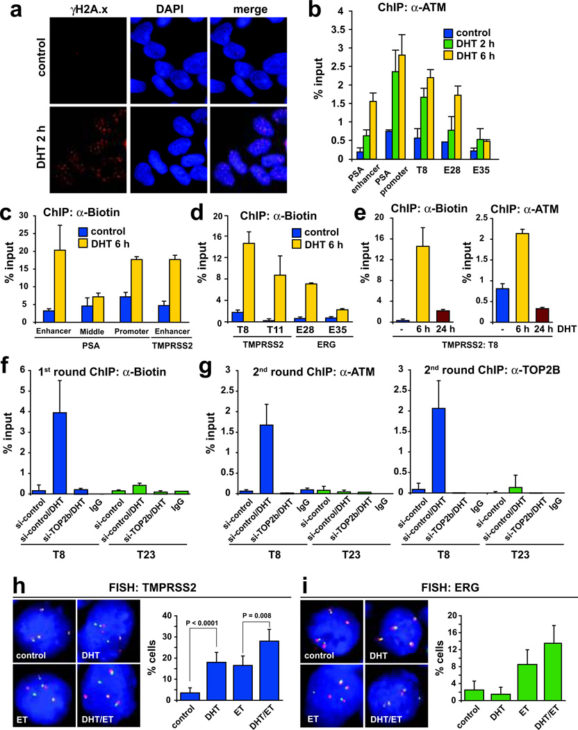
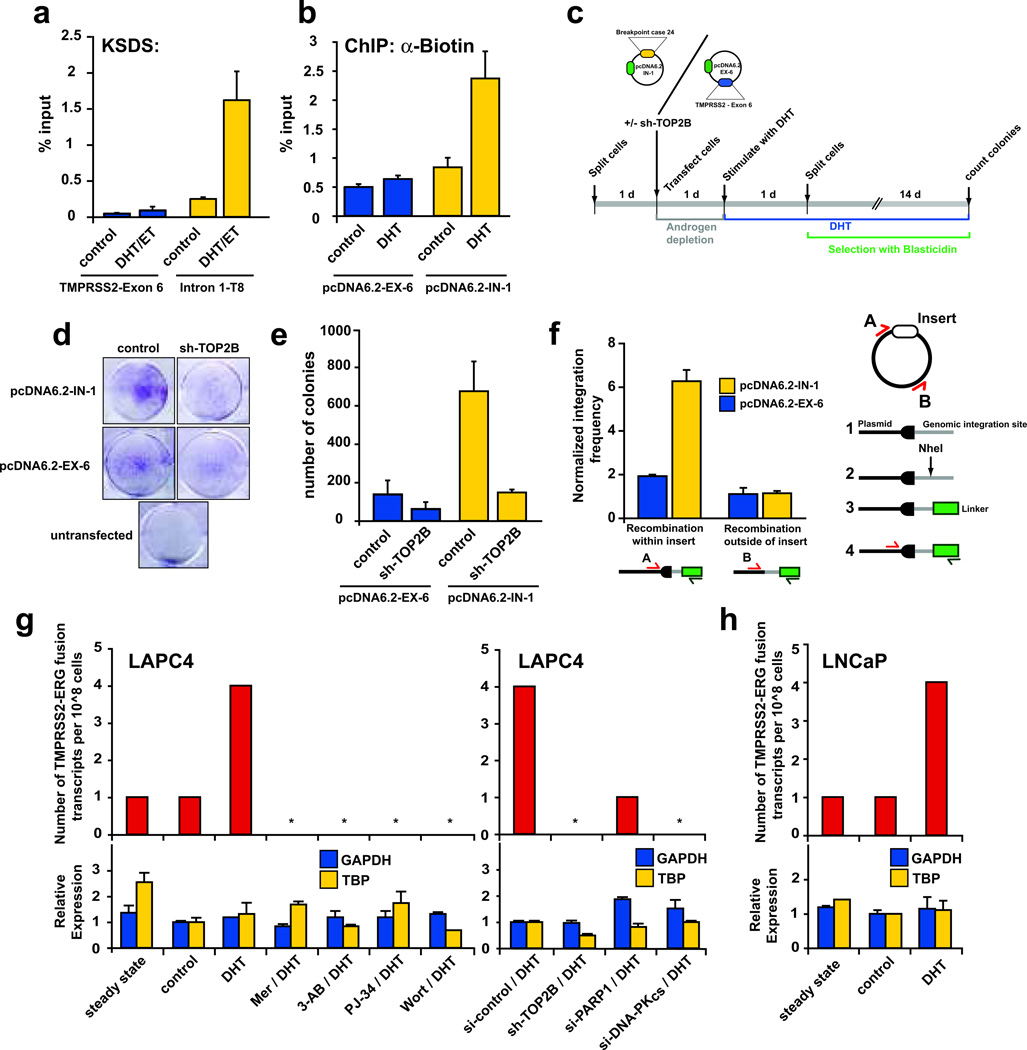
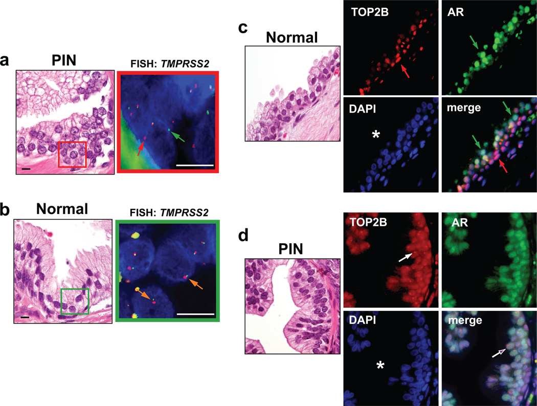
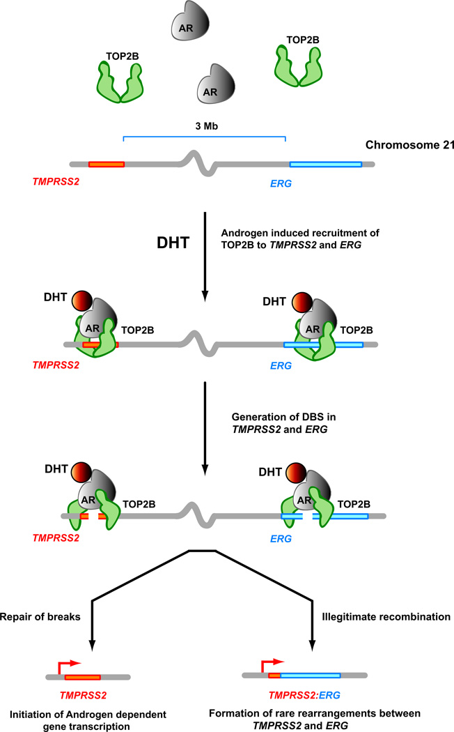
DNA double-strand breaks (DSBs) can lead to the development of genomic rearrangements, which are hallmarks of cancer. Fusions between TMPRSS2, encoding the transmembrane serine protease isoform 2, and ERG, encoding the v-ets erythroblastosis virus E26 oncogene homolog, are among the most common oncogenic rearrangements observed in human cancer. We show that androgen signaling promotes co-recruitment of androgen receptor and topoisomerase II beta (TOP2B) to sites of TMPRSS2-ERG genomic breakpoints, triggering recombinogenic TOP2B-mediated DSBs. Furthermore, androgen stimulation resulted in de novo production of TMPRSS2-ERG fusion transcripts in a process that required TOP2B and components of the DSB repair machinery. Finally, unlike normal prostate epithelium, prostatic intraepithelial neoplasia cells showed strong coexpression of androgen receptor and TOP2B. These findings implicate androgen-induced TOP2B-mediated DSBs in generating TMPRSS2-ERG rearrangements.


Figures

Figure 1
Figure 1
TOP2B is required for efficient induction of AR target gene expression following androgen stimulation. a, Inhibition of TOP2B with ET or Mer prior to DHT stimulation of LNCaP cells attenuated expression of an androgen-induced geneset (p < 10−14), defined as the 45 genes upregulated by >2-fold in DHT vs. control treated LNCaP cells. b, Geneset enrichment analysis shows highly statistically significant enrichment for down regulation of three independently derived but overlapping androgen-responsive genesets in ET/DHT vs. DHT, Mer/DHT vs. DHT, and sh-TOP2B/DHT vs DHT (see also Supplementary Fig. 2,3). Y-axis shows the degree of confidence for geneset enrichment as −log10(p-value). Significance threshold corresponding to p = 0.05 is indicated. c–d, Real-time RT-PCR analysis of relative expression of selected genes with respect to GAPDH expression confirms down-regulation of representative androgen-responsive genes in ET/DHT, Mer/DHT, and sh-TOP2B/DHT. Data are shown as mean ± SE of two to three replicates.
Figure 2
Figure 2
Androgen stimulation induced co-recruitment of AR/TOP2B and TOP2B catalytic cleavage at known regulatory regions of AR target genes. a, schematic of AREs in the promoter and enhancer region of PSA and TMPRSS2. Bars indicate positions (relative to transcriptional start site) of amplicons analyzed. b, ChIP analysis of androgen-deprived LAPC4 cells that were stimulated with 100 nM DHT for varying timepoints as indicated reveals DHT-induced recruitment of AR and TOP2B to the enhancer and promoter of PSA and TMPRSS2 but not to an intervening region (middle) between the enhancer and promoter of PSA. c, Stimulation of androgen-depleted LAPC4 cells with 100 nM DHT for 6 h allows co-immunoprecipitation of TOP2B with AR antibodies and vice versa. IP, immunoprecipitating antibody; IB, immunoblotting antibody; TCL, total cell lysate. d, ChIP-re-ChIP experiments demonstrate that stimulation of androgen-deprived LAPC4 cells with 100 nM DHT induces co-recruitment of AR and TOP2B to AREs of PSA and TMPRSS2. A first round of ChIP was performed using anti-TOP2B antibodies and the resulting immunoprecipitates were subjected to a second round of ChIP with anti-AR antibodies. Relative enrichment was determined by qPCR and is shown as percentage of input DNA. e–f, KSDS method shows TOP2 catalytic cleavage of AR target sites in LAPC4 cells in a DHT and TOP2B dependent fashion. Results are presented as mean percentage to input with ± SE of two to three replicates.
Figure 3
Figure 3
Androgen stimulation induces AR/TOP2B recruitment and TOP2B catalytic cleavage at genomic breakpoints of TMPRSS2 and ERG observed in human PCa. a–b, Sites closest to TMPRSS2-ERG breakpoints from 8 PCa cases determined from various studies (arrows) were significantly enriched for high KSDS enrichment of TOP2 catalytic cleavage (p = 0.010 and 0.013 respectively) in LAPC4 cells. Labeled sites (e.g., T8, T23, E5, E13, etc.) are analyzed in subsequent experiments. c, DHT and TOP2B dependent TOP2 catalytic cleavage in LAPC4 cells around the case 24 breakpoint aligning with region T8 (upper panel). The lack of KSDS enrichment at region E47 at ERG was re-confirmed in an independent KSDS experiment (lower panel). d, SLOT assay showed that DHT-induced TOP2 catalytic activity in LAPC4 cells was significantly higher at an NspI fragment most proximal to the TMPRSS2 breakpoint observed in case 24 than to the adjacent, more distal NspI fragment (see Supplementary Fig. 8, 9 for overview of SLOT). e, ChIP enrichment of AR and TOP2B at representative TOP2 catalytic cleavage sites in DHT-stimulated LAPC4 cells relative to untreated controls. f–g, 3C analysis reveals DHT-dependent spatial chromatin interaction of TMPRSS2 enhancer and promoter with region T8 (see first lane vs. fourth lane). Omission of NspI restriction enzyme and/or DNA ligase served as assay negative controls. Inhibition with Mer prevented these DHT-induced interactions. Error bars indicate ± SE of two to three experiments.
Figure 4
Figure 4
Androgen stimulation results in TOP2B dependent DSB formation. a, Androgen stimulation induced γH2A.x foci formation in LAPC4 cells. b, Androgen stimulation led to recruitment of ATM to representative sites of high TOP2B activity but much less so to a region showing low TOP2B catalytic cleavage (E35). c–d, Stimulation of LAPC4 cells with DHT induced DSB that could be end-labeled with biotin at representative TOP2 cleavage sites, but not to regions showing low TOP2 cleavage (PSA middle, E35). e, DSB largely resolve by 24 h following DHT stimulation of LAPC4 cells as evidenced by reduced biotin labeling and ATM recruitment. The time course of DSB formation/resolution and ATM recruitment are highly parallel. f, Left panel, DHT-induced DSB at a representative TOP2B enriched site (T8) are dependent on TOP2B. A site showing low TOP2B enrichment (T23) served as a negative control. g, DHT-induced strand breaks at representative TMPRSS2 sites are enriched for TOP2B and ATM binding as seen by ChIP-re-ChIP experiments. Results are presented as mean percentage to input with ± SE of two to three replicates. h, DHT induces chromosomal breaks at TMPRSS2 in LAPC4 cells, monitored as fraction of cells (n > 200) showing “split-apart” of FISH probes 5’ (green) and 3’ (red) of TMPRSS2. i, Treatment of LAPC4 cells with ET plus/minus DHT increased split-aparts at ERG compared to control or DHT treatment. Error bars indicate 95% confidence intervals.
Figure 5
Figure 5
Androgen-induced TOP2B mediated DSB are recombinogenic and promote de novo production of TMPRSS2-ERG fusion genes. a, Selection of TMPRSS2 regions showing high (Intron 1-T8) and low (TMPRSS2-Exon 6) KSDS enrichment in response to DHT stimulation of LAPC4 cells. b, DHT-induced breaks can be detected in plasmids containing sequences surrounding region T8 of TMPRSS2 intron 1 (pcDNA6.2-IN-1) as evidenced by increased biotin labeling in DHT stimulated LAPC4 cells transfected with this plasmid. Plasmids containing TMPRSS2 exon 6 (pcDNA6.2-EX-6) served as a negative control. c, Schematic of androgen-induced genomic recombination assay in LAPC4 cells transfected with pcDNA6.2-IN-1 or pcDNA6.2-EX-6, which contain a blasticidin resistance gene. Number of colonies represents the number of recombination events allowing integration of the blasticidin resistance vectors into the LAPC4 genome. d, Representative results of genomic recombination assays. e, pcDNA6.2-IN-1 transfected LAPC4 cells produced significantly more androgen induced recombination events than pcDNA6.2-EX-6 transfected cells. Treatment with sh-TOP2B abolished this effect. f, While both showed similar recombination frequency at the vector backbone, recombination frequency within the IN-1 insert was significantly higher than that in the EX-6 insert in pooled colonies as determined using the strategy shown on the right. Data are shown as mean ± SE of two to three replicates. g, DHT-stimulation of LAPC4 cells leads to increased TMPRSS2-ERG fusion transcripts compared to background levels in LAPC4 cells grown in androgen-containing (steady-state) or androgen-deprived (control) media. Pharmacological or genetic modulation of TOP2B (Mer, sh-TOP2B), PARP1 (3-AB, PJ-34, si-PARP1), or DNA-PKCS (Wort, si-DNA-PKCS), reduces TMPRSS2-ERG fusion transcripts without significantly altering GAPDH or TBP expression. h, DHT-stimulation leads to de novo formation of TMPRSS2-ERG fusion transcripts in LNCaP cells.
Figure 6
Figure 6
TMPRSS2-ERG rearrangements are observed in PIN PCa precursor lesions and are associated with changes in the TOP2B expression pattern. a, H&E stain and corresponding TMPRSS2 FISH in a representative PIN lesion. FISH image of red boxed area shows interphase nuclei of PIN lesion with split-aparts of 5’ (green arrow) and 3’ (red arrow) TMPRSS2 FISH probes. b, H&E stain and corresponding TMPRSS2 FISH (green boxed area) of normal prostate epithelium. Orange arrows indicate normal configuration of 5’ and 3’ TMPRSS2 FISH probes. Scale bars indicate 10 µm c, Normal prostate epithelium shows strong expression of TOP2B in basal cells (red arrow) and low expression in luminal cells, whereas AR is predominantly expressed in luminal cells (green arrow). Lumen of prostate gland is marked with an asterisk. d, PIN lesion shows high TOP2B in basal and luminal cells (purple arrow). Note the accentuated nucleolar localization of TOP2B (white arrow).
Figure 7
Figure 7
A proposed model for androgen-induced TOP2B mediated double strand breaks and TOP2B instability (TIN) for the generation of DSB and formation of TMPRSS2-ERG gene fusions. Androgen stimulation leads to co-recruitment of liganded AR and TOP2B to regulatory regions of androgen responsive genes as well as to regions of TMPRSS2 and ERG genes observed to participate in genomic rearrangements. TOP2B catalytic activity and the associated DSB formation is required for efficient activation of androgen responsive genes, and these DSB are usually resolved shortly after induction. In some circumstances, stabilization of these TOP2B-mediated DSB can lead to illegitimate recombination and rare rearrangements between TMPRSS2 or other androgen responsive genes and ERG that are then subject to selection during neoplastic outgrowth.

Comment in

  • On the origin of prostate fusion oncogenes.
    Bartek J, Hamerlik P, Lukas J. Bartek J, et al. Nat Genet. 2010 Aug;42(8):647-8. doi: 10.1038/ng0810-647. Nat Genet. 2010. PMID: 20664645 No abstract available.
  • Prostate cancer: Gene fusions.
    Groves-Kirkby N. Groves-Kirkby N. Nat Rev Urol. 2010 Sep;7(9):473. doi: 10.1038/nrurol.2010.125. Nat Rev Urol. 2010. PMID: 20836279 No abstract available.

References

    1. Rowley JD. Chromosomal translocations: revisited yet again. Blood. 2008;112:2183–2189. - PubMed
    1. Mitelman F, Johansson B, Mertens F. The impact of translocations and gene fusions on cancer causation. Nat Rev Cancer. 2007;7:233–245. - PubMed
    1. Morgan WF, et al. DNA double-strand breaks, chromosomal rearrangements, and genomic instability. Mutat Res. 1998;404:125–128. - PubMed
    1. Richardson C, Jasin M. Frequent chromosomal translocations induced by DNA double-strand breaks. Nature. 2000;405:697–700. - PubMed
    1. Kumar-Sinha C, Tomlins SA, Chinnaiyan AM. Recurrent gene fusions in prostate cancer. Nat Rev Cancer. 2008;8:497–511. - PMC - PubMed

References for Methods

    1. Boonyaratanakornkit V, et al. High-mobility group chromatin proteins 1 and 2 functionally interact with steroid hormone receptors to enhance their DNA binding in vitro and transcriptional activity in mammalian cells. Mol Cell Biol. 1998;18:4471–4487. - PMC - PubMed
    1. Wang Q, Carroll JS, Brown M. Spatial and temporal recruitment of androgen receptor and its coactivators involves chromosomal looping and polymerase tracking. Mol Cell. 2005;19:631–642. - PubMed
    1. Splinter E, Grosveld F, de Laat W. 3C technology: analyzing the spatial organization of genomic loci in vivo. Methods Enzymol. 2004;375:493–507. - PubMed
    1. Spitzner JR, Chung IK, Muller MT. Eukaryotic topoisomerase II preferentially cleaves alternating purine-pyrimidine repeats. Nucleic Acids Res. 1990;18:1–11. - PMC - PubMed
    1. DeWeese TL, et al. Human papillomavirus E6 and E7 oncoproteins alter cell cycle progression but not radiosensitivity of carcinoma cells treated with low-dose-rate radiation. Int J Radiat Oncol Biol Phys. 1997;37:145–154. - PubMed

Publication types