. 2010 Jul;17(7):882-8.
doi: 10.1038/nsmb.1837. Epub 2010 Jul 4.

Crystal structure of the conserved herpesvirus fusion regulator complex gH-gL

Affiliations

Crystal structure of the conserved herpesvirus fusion regulator complex gH-gL

Tirumala K Chowdary et al. Nat Struct Mol Biol. 2010 Jul.

Abstract

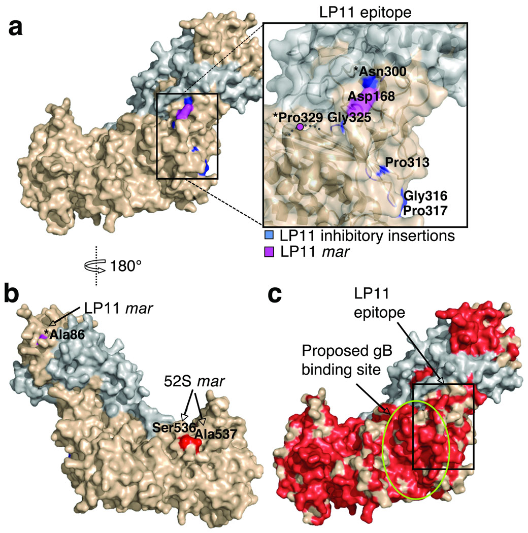
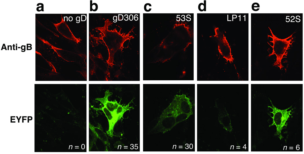
Herpesviruses, which cause many incurable diseases, infect cells by fusing viral and cellular membranes. Whereas most other enveloped viruses use a single viral catalyst called a fusogen, herpesviruses, inexplicably, require two conserved fusion-machinery components, gB and the heterodimer gH-gL, plus other nonconserved components. gB is a class III viral fusogen, but unlike other members of its class, it does not function alone. We determined the crystal structure of the gH ectodomain bound to gL from herpes simplex virus 2. gH-gL is an unusually tight complex with a unique architecture that, unexpectedly, does not resemble any known viral fusogen. Instead, we propose that gH-gL activates gB for fusion, possibly through direct binding. Formation of a gB-gH-gL complex is critical for fusion and is inhibited by a neutralizing antibody, making the gB-gH-gL interface a promising antiviral target.


Figures

Fig. 1
Fig. 1. Structure of HSV-2 gH–gL complex
(a) Domain arrangement. Regions present in the expression construct but missing from the final model are shown as dashed lines. gH domains are in green (H1), yellow (H2) and red (H3); gL is in blue. The numbering scheme for gH cysteines is based on HSV-1 gH, which has eight cysteines. HSV-2 gH has seven cysteines, lacking C3. (b) Side view of gH–gL structure showing disordered segments (dotted lines), disulfides (yellow spheres and red sticks), and sugars (grey). (c) gH–gL in molecular surface representation. View in the top panel is the same as in (b). View in the bottom panel was generated by 180° rotation around the vertical axis.
Fig. 2
Fig. 2. Domains of gH and gL
(a) domain H1, (b) domain H2, (c) domain H3, (d) gL. The coloring scheme is the same as in Fig. 1. Disulfides are shown as yellow spheres and red sticks and labeled. All secondary structure elements are labeled. Disordered segments are shown as dotted lines. Labeled residues indicate the limits of individual domains and the disordered loops.
Fig. 3
Fig. 3. gH–gL interface
(a) The gH “clamp”. The molecular surface of gH is shown in beige; gL is rendered as a blue ribbon. Inset: up-close view of the H1/gL interface. Secondary structure at the H1/gL interface is labeled. (b) “Open-book” view of the H1A/gL interface viewed from the “heel” of the gH–gL “boot”. (c) “Open-book” view of the H1B/gL interface viewed from the top of the gH–gL “boot”. Intermolecular interactions in (b) and (c) are color-coded as follows: buried hydrophobic surface (light blue), buried hydrophilic surface (green), and negatively charged (red) and positively charged (blue) residues, in salt bridges.
Fig. 4
Fig. 4. Locations of several predicted heptad repeats and fusion peptides in gH
(a) predicted heptad repeat sequences in gH that inhibit cell-cell fusion as synthetic peptides, residues 444–479 and 542–582, are shown in purple and labeled. Domain H3 is shown as a light pink surface. (b) putative fusion peptide sequences of gH, residues 626–644 and 766–797, are shown in purple and labeled. Domain H3 is shown as light pink ribbon. Secondary structure elements within domains H2 and H3 are numbered according to Figs. 2 and 3.
Fig. 5
Fig. 5. The epitope of the HSV-1 neutralizing antibody LP11 defines the gB-binding site
(a) gH–gL in surface representation, gH (beige) and gL (grey). The proposed LP11 epitope is boxed and enlarged in the inset. (b) gH–gL is color-coded as in (a). The view in (b) is rotated 180° around the vertical axis relative to (a). Residues with a star are not conserved between HSV-1 and HSV-2. (c) gH–gL is colored by sequence conservation between HSV-1 and HSV-2, with identical residues in red. The view is as in (a). LP11 epitope is boxed. mar is monoclonal antibody resistance mutation.
Fig. 6
Fig. 6. Effect of anti-gH antibodies on gB–gH BiMC
Top panels show gB surface expression (red) detected using immunofluorescence. Bottom panels show EYFP BiMC (bright green fluorescence). (a) no gD, (b) gD306, (c) gD306 plus mAb 53S, (d) gD306 plus mAb LP11, and (e) gD306 plus mAb 52S. n is the number of syncytia from a representative experiment. EYFP is enhanced yellow fluorescent protein.

Similar articles

Cited by

References

    1. Spear PG, Longnecker R. Herpesvirus entry: an update. J Virol. 2003;77:10179–10185. - PMC - PubMed
    1. Heldwein EE, Krummenacher C. Entry of herpesviruses into mammalian cells. Cell Mol Life Sci. 2008;65:1653–1668. - PMC - PubMed
    1. Ryckman BJ, et al. Characterization of the human cytomegalovirus gH/gL/UL128-131 complex that mediates entry into epithelial and endothelial cells. J Virol. 2008;82:60–70. - PMC - PubMed
    1. Wickner W, Schekman R. Membrane fusion. Nat Struct Mol Biol. 2008;15:658–664. - PMC - PubMed
    1. Heldwein EE, et al. Crystal structure of glycoprotein B from herpes simplex virus 1. Science. 2006;313:217–220. - PubMed

Publication types

Associated data