Review
. 2010 Oct;67(20):3467-88.
doi: 10.1007/s00018-010-0437-0. Epub 2010 Jul 4.

Molecular aspects of cyclophilins mediating therapeutic actions of their ligands

Affiliations
Review

Molecular aspects of cyclophilins mediating therapeutic actions of their ligands

Andrzej Galat et al. Cell Mol Life Sci. 2010 Oct.

Abstract

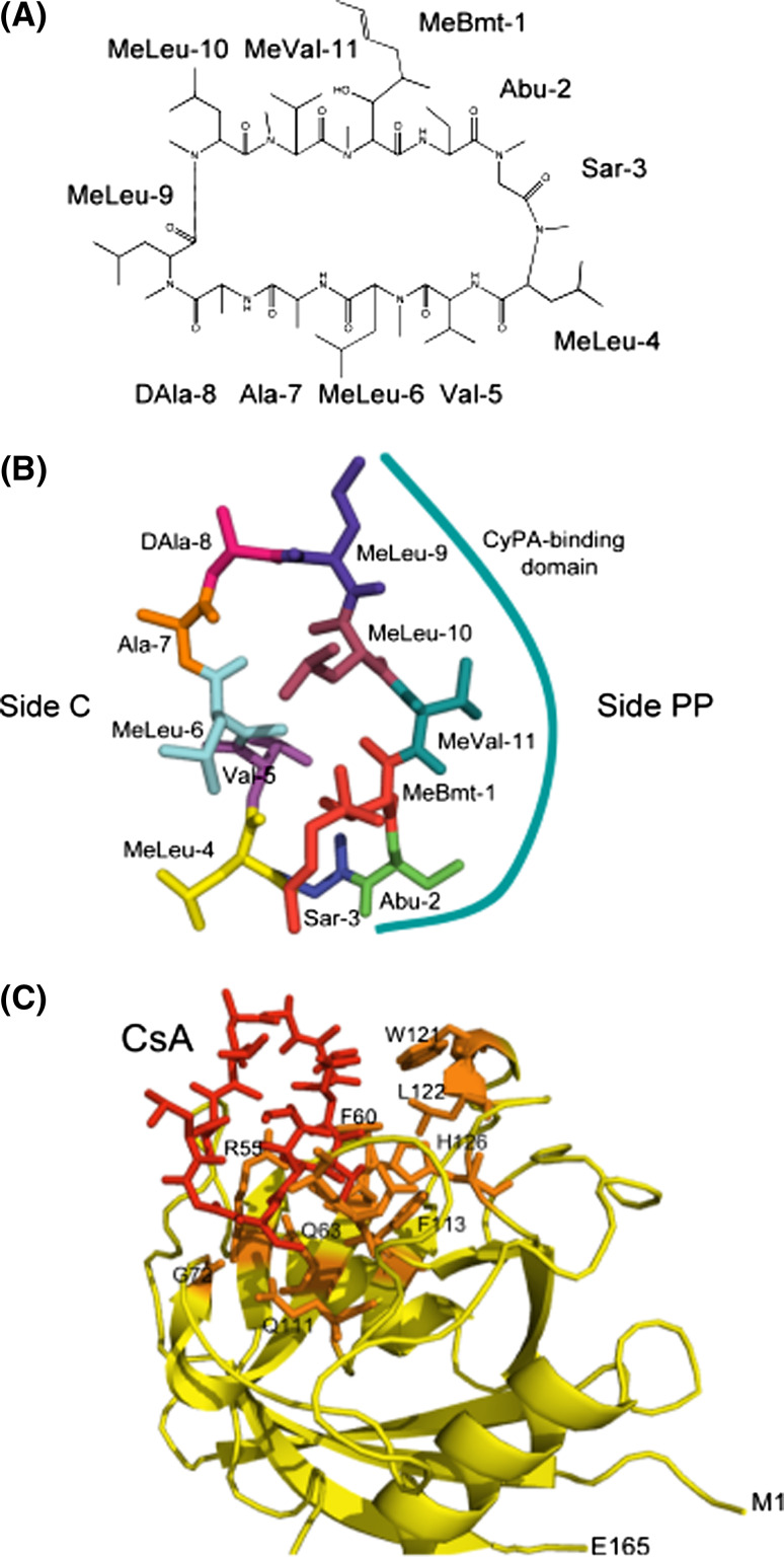
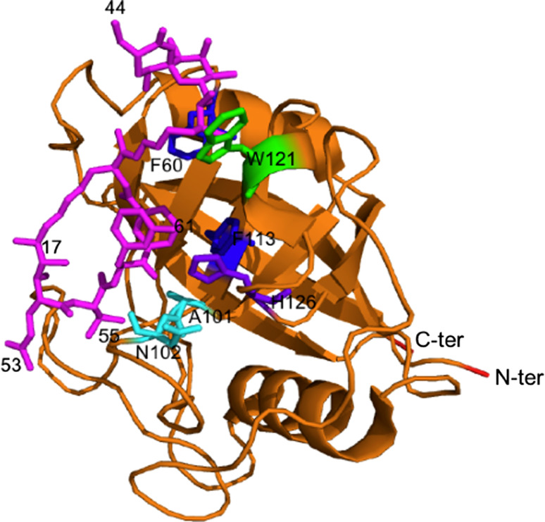
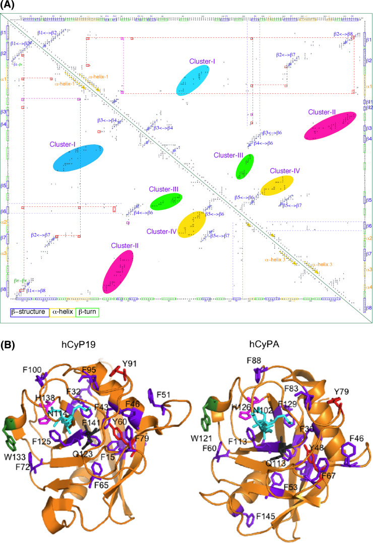
Cyclosporine A (CsA) is an immunosuppressive cyclic peptide that binds with a high affinity to 18 kDa human cyclophilin-A (hCyPA). CsA and its several natural derivatives have some pharmacological potential in treatment of diverse immune disorders. More than 20 paralogues of CyPA are expressed in the human body while expression levels and functions of numerous ORFs encoding cyclophilin-like sequences remain unknown. Certain derivatives of CsA devoid of immunosuppressive activity may have some potential in treatments of Alzheimer diseases, Hepatitis C and HIV infections, amyotrophic lateral sclerosis, congenital muscular dystrophy, asthma and various parasitic infections. Here, we discuss structural and functional aspects of the human cyclophilins and their interaction with various intra-cellular targets that can be under the control of CsA or its complexes with diverse cyclophilins that are selectively expressed in different cellular compartments. Some molecular aspects of the cyclophilins expressed in parasites invading humans and causing diseases were also analyzed.


Figures

Fig. 1
Fig. 1
Cyclosporine-A. a The chemical structure of CsA with the following abbreviations: Abu L-α-aminobutiric acid, MeBmt (4R)-4[(E)-2-butenyl]-4,N-dimethyl-L-threonine, MeLeu N-methylleucine, MeVal N-methylvaline, Sar sarcosine. b CsA structure extracted from its complex with hCyPA (1CWA.pdb); the side interacting with CyPA (side PP) is marked with the solid line and side C with the residues interacting with calcineurin. c Full structure of the hCyPA/CsA complex (1CWA.pdb); CsA is marked as red sticks, the AA residues forming the PPIase cavity are shown as orange sticks while the remaining backbone of the cyclophilin is in yellow. M1 N-terminal methionine, E165 C-terminal glutamic acid
Fig. 2
Fig. 2
Sanglifehrin A (pink sticks) bound to hCyPA (ribbon structure made from 1YND.pdb [52])
Fig. 3
Fig. 3
Information entropy (I e) histogram for the human CLDs (see ESM Fig. Fs5). Low I e values indicate that thye given sequence position is well conserved (a low variability in the AAC) whereas the I e ≥ 1.0 indicate for a higher variability of the AAC at the given sequence position (maximal I e for 20 natural AAs equals to log220 = 4.322)
Fig. 4
Fig. 4
a 2D map of intra-molecular atomic distances calculated in the range 2.7–4.5 Å in the structures of hCyP19 [18] (1QOI.pdb, upper triangle) and hCyPA (2CPL.pdb, lower triangle) [16]. Integer numbers correspond to the sum of the number of inter-residues distances (NIDs) for given pairs of AA residues. At the upper axis and the right side is shown the sequence of hCyP19, whereas the bottom axis has the sequence of hCyPA. b X-ray structures of hCyP19 (1QOI.pdb, left panel), and hCyPA (2CPL.pdb, right panel); the spatial positions of F (violet), Y (red), W (deep green), catalytically crucial MAN motif, E111 (hCyPA) and H126 (hCyPA) are indicated in cyan, black and pink sticks, respectively. The ribbon structures were made with the PyMol program [61]; see Fs4A in ESM
Fig. 5
Fig. 5
Numbers of interactions (upper panel) and the sum of vdW and Coulombic energy terms [19] per each residue (lower panel) calculated from the X-ray structure of hCyP19 (1QOI.pdb) with written patches of sequences that are crucial for the PPIase cavity and the aromatic/hydrophobic AAs network; red dashed line was arbitrarily placed to indicate the largest energy contributions. The upper parts of the sequence patches come from hCyP19 whereas lower parts were taken from the sequence of hCyPA
Fig. 6
Fig. 6
Three X-ray structures of the binary and tertiary complexes; a the (hCyPA/CsA + CnA/CnB) complex [13]; CyPA (yellow) binds to CsA (red, indicated with a black arrow) and interacts with CnA (orange ribbons) and CnB (green ribbon) complex. b X-ray structure of HIV-1 capsid protein (violet) bound to the PPIase cavity of hCyPA (yellow ribbon); the loop in red comes from the capsid protein (1M9C.pdb, [30]). c X-ray structure of a short peptide from the U4/U6 snRNP-60K (violet) protein interacting with the hCyP19 (yellow ribbon) with some of the AAs forming its PPIase cavity (red sticks) (1MZW.pdb, [18])
Fig. 7
Fig. 7
The human cyclophilins and their respective orthologues encoded in the genomes of Trypanosoma cruzi, Plasmodium falciparum and Toxoplasma gondii. The proteins were arranged according to two criteria, namely (1) presence of similar domains, and (2) similar sequence attributes for their respective CLDs in the cases where the large cyclophilins were aligned with their small homologues
Fig. 8
Fig. 8
Chemical structures of derivatives of CsA devoid of immunosuppressive activity in humans. Continuous lines indicate the places where chemical modifications were made while stripped line on CsA indicates the residues that should be hidden inside the PPIase cavity
Fig. 9
Fig. 9
Distribution of hydrophobicity versus pI of the human CLDs (see Table 2)

Similar articles

Cited by

References

    1. Borel JF, Feurer C, Gubler HU, Stahelin H. Biological effects of cyclosporine A: a new antilymphocytic agent. Agents Actions. 1994;43:179–186. - PubMed
    1. Handschumacher RE, Harding MW, Rice J, Drugge RJ, Speicher DW. Cyclophilin: a specific cytosolic binding protein for cyclosporine A. Science. 1984;226:544–546. - PubMed
    1. Galat A, Riviere S. Peptidylprolyl cis/trans isomerases. Oxford: Oxford University Press; 1998.
    1. Edlich F, Fischer G. Pharmacological targeting of catalized protein folding: the example of peptide bond cis/trans isomerases. Hand Exp Pharmacol. 2006;172:359–404. - PubMed
    1. Galat A, Bouet F. Cyclophilin B is an abundant protein whose conformation is similar to that of cyclophilin A. FEBS Lett. 1994;347:31–36. - PubMed

Publication types

LinkOut - more resources