. 2010 Aug;27(8):583-96.
doi: 10.1002/yea.1797.

A screen for deficiencies in GPI-anchorage of wall glycoproteins in yeast

Affiliations

A screen for deficiencies in GPI-anchorage of wall glycoproteins in yeast

Marlyn Gonzalez et al. Yeast. 2010 Aug.

Abstract

Many of the genes and enzymes critical for assembly and biogenesis of yeast cell walls remain unidentified or poorly characterized. Therefore, we designed a high throughput genomic screen for defects in anchoring of GPI-cell wall proteins (GPI-CWPs), based on quantification of a secreted GFP-Sag1p fusion protein. Saccharomyces cerevisiae diploid deletion strains were transformed with a plasmid expressing the fusion protein under a GPD promoter, then GFP fluorescence was determined in culture supernatants after mid-exponential growth. Variability in the amount of fluorescent marker secreted into the medium was reduced by growth at 18 degrees C in buffered defined medium in the presence of sorbitol. Secondary screens included immunoblotting for GFP, fluorescence emission spectra, cell surface fluorescence, and cell integrity. Of 167 mutants deleted for genes affecting cell wall biogenesis or structure, eight showed consistent hyper-secretion of GFP relative to parental strain BY4743: tdh3 (glyceraldehyde-3-phosphate dehydrogenase), gda1 (guanosine diphosphatase), gpi13 and mcd4 (both ethanolamine phosphate-GPI-transferases), kre5 and kre1 (involved in synthesis of beta1,6 glucan), dcw1(implicated in GPI-CWP cross-linking to cell wall glucan), and cwp1 (a major cell wall protein). In addition, deletion of a number of genes caused decreased secretion of GFP. These results elucidate specific roles for specific genes in cell wall biogenesis, including differentiating among paralogous genes.


Figures

Figure 1
Figure 1
Cell wall localization of GFP–Sag1p. (A) Schematic of pGFP–Sag1p reporter construct in low copy vector p416-GPD. (B1) Immunoelectron micrographs of wild-type cells expressing GFP–Sag1p labelled with mouse monoclonal anti-GFP as primary antibody and gold-labelled goat anti-mouse as secondary antibody. (B2) Immunoelectron micrograph of non-expressing cells. (C) Phase contrast and GFP fluorescence of isolated washed cell walls from BY4743 cells expressing GFP–Sag1p (C1) and BY4743 cells transformed with empty vector alone (C2). Cell surface fluorescence of: (D1) BY4743–GFP–Sag1p wild-type; (D2) KRE5/kre5 mutant; and (D3) non-expressing cells
Figure 2
Figure 2
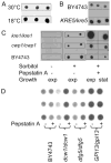
Immunoblot analyses of cell wall mutants and optimization of assays. (A) Growth at lower temperature increases GFP–Sag1p yields. Triplicate clones of BY4743 were grown at 30 °C (top) and 18 °C before assay. (B) Supernatants from triplicate clones of BY4743 (top) and KRE5/kre5 mutant grown without pepstatin A. (C) Effect of sorbitol (1 M), pepstatin A (1 μM) and growth phase. Duplicate clones were grown without sorbitol or with sorbitol in the absence (left) or presence (right) of pepstatin A. (D) Effect of growth in pepstatin A (1 μM) on secretion of GFP–Sag1p in wild-type and three deletion strains. Each strain was grown in triplicate under standard conditions
Figure 3
Figure 3
Supernatant fluorescence values for representative mutants. Mutant strains were grown without pepstatin A in triplicate at 18 °C to mid-log phase in osmotically stable medium buffered to pH 6.5, and supernatants were assayed for GFP fluorescence. Mutant names and genotypes are given in Table S2 (see Supporting information). Mean and SEM values (dashed lines) are shown for the wild-type BY4743–GFP–Sag1 strain and for mutant strains (error bars)
Figure 4
Figure 4
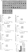
Confirmation of hyper- and hyposecreting mutants. (A) Standard condition immunoblots of growth supernatants from five replicate clones of BY4743 and seven mutants. (B1) Uncorrected spectra for GFP and culture supernatant of BY4743. (B2) corrected spectrum for BY4743. (B3–B7) Background-corrected fluorescence spectra from supernatants from BY4743 (dashed lines), selected mutants (dotted lines) and purified GFP (solid lines) as reference. The relative fluorescence for each sample was taken as the height at the maximum point in the corrected spectrum (C). Cells were grown without pepstatin A
Figure 5
Figure 5
Cell surface GFP fluorescence of BY4743 and selected mutants. Fluorescence exposure series for wild-type (A), MCD4/mcd4 (B) and GPI13/gpi13 (C). Also shown are details of septum regions for MCD4/mcd4 (D) and GPI13/gpi13 (E). On the right are matched exposures of BY4743 (F), gda1/gda1 (G) and tdh3/tdh3 (H). At the bottom are matched exposures of (I) BY4743, (J) dcw1/dcw1 and (K) dfg5/dfg5

References

    1. Aanen DK, Slippers B, Wingfield MJ. Biological pest control in beetle agriculture. Trends Microbiol. 2009;17:179–182. - PubMed
    1. Abeijon C, Yanagisawa K, Mandon EC, et al. Guanosine diphosphatase is required for protein and sphingolipid glycosylation in the Golgi lumen of Saccharomyces cerevisiae. J Cell Biol. 1993;122:307–323. - PMC - PubMed
    1. Adesemoye AO, Kloepper JW. Plant–microbes interactions in enhanced fertilizer-use efficiency. Appl Microbiol Biotechnol. 2009;85:1–12. - PubMed
    1. Alloush HM, Lopez-Ribot JL, Masten BJ, et al. 3-Phosphoglycerate kinase: a glycolytic enzyme protein present in the cell wall of Candida albicans. Microbiology. 1997;143(2):321–330. - PubMed
    1. Almeida AM, Hillmen P, Richards SJ, et al. Defective modification of mannose residues by terminal phosphoethanolamine underlies inherited GPI deficiency. J Am Soc Hematol. 2005;106 abstr 128.

Publication types

MeSH terms

Substances