. 2010 Jun 25;141(7):1230-40.
doi: 10.1016/j.cell.2010.05.007.

Arabidopsis HEMERA/pTAC12 initiates photomorphogenesis by phytochromes

Affiliations

Arabidopsis HEMERA/pTAC12 initiates photomorphogenesis by phytochromes

Meng Chen et al. Cell. .

Abstract

Light plays a profound role in plant development, yet how photoreceptor excitation directs phenotypic plasticity remains elusive. One of the earliest effects of light is the regulated translocation of the red/far-red photoreceptors, phytochromes, from the cytoplasm to subnuclear foci called phytochrome nuclear bodies. The function of these nuclear bodies is unknown. We report the identification of hemera, a seedling lethal mutant of Arabidopsis with altered phytochrome nuclear body patterns. hemera mutants are impaired in all phytochrome responses examined, including proteolysis of phytochrome A and phytochrome-interacting transcription factors. HEMERA was identified previously as pTAC12, a component of a plastid complex associated with transcription. Here, we show that HEMERA has a function in the nucleus, where it acts specifically in phytochrome signaling, is predicted to be structurally similar to the multiubiquitin-binding protein, RAD23, and can partially rescue yeast rad23mutants. Together, these results implicate phytochrome nuclear bodies as sites of proteolysis.


Figures
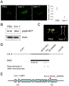
Figure 1
Figure 1. Isolation and map-based cloning of hmr
(A) Confocal images showing subnuclear localization of PHYB∷GFP in epidermal cells at the top of the hypocotyl of PBG and hmr-1 under 8 μmol m-2 sec-1 of R light. PHYB∷GFP was localized to large NBs with an average diameter of 1.6 μm in PBG, whereas PHYB∷GFP NBs in hmr-1 were smaller, with an average diameter of 0.4 μm. In a small fraction of hmr-1 hypocotyl cells, PHYB∷GFP was evenly dispersed in the nucleoplasm. (B) Protein levels of PHYB∷GFP remained the same in PBG and hmr-1 seedlings. Total protein extracts from 4-day R light grown hmr-1 and PBG seedlings were resolved by SDS-PAGE. Protein levels of PHYB∷GFP were detected by western blot using anti-GFP antibodies. Actin was used as a loading control. (C) Images of 4-day old PBG and hmr-1 seedlings grown under 8 μmol m-2 sec-1 of R light. The hmr-1 mutant was taller compared to PBG. (D) Map-based cloning of hmr. The hmr-1 mutation was mapped to chromosome II on BAC T31E10 between markers MC671672 and MC549550 based on an F2 mapping population of 1960 plants generated by crossing hmr-1 (Ler) and Col-0. The interval contains five predicted genes illustrated as arrows. The bold arrow represents the HMR gene, At2g34640. (E) Schematic illustration of the exon-intron structure of HMR with the shaded boxes representing exons. The mutations in hmr-1 and hmr-2 are indicated by red arrows. See also Figure S1.
Figure 2
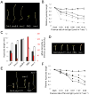
Figure 2. hmr mutants are defective in multiple PHYB- and PHYA-mediated responses
(A) Images of 4-day old Col-0, phyB-9, hmr-2, PBG, and hmr-1 seedlings grown in 8 μmol m-2 sec-1 R light. (B) Fluence response curves for Rc. Relative hypocotyl length of 4-day grown Col-0 (◆), phyB-9 (formula image), hmr-2 (●), PBG (formula image), hmr-1 (formula image), and phyB-5 (formula image) seedlings under different fluence of Rc and dark conditions. (C) EOD-FR responses of Col-0, hmr-2, phyB-9, PBG, and hmr-1. Filled columns represent hypocotyl lengths of 4-day old seedlings under 8-hour day/16-hour night; open columns represent hypocotyl lengths of 4-day old seedlings under the same short day conditions with an additional 15 min FR treatment at the end of the day. Red columns represent the percentage of increase in hypocotyl length of the treated seedlings compared to untreated seedlings. (D) Cotyledon opening responses for VLFR measurement. Cotyledon images of Col-0, phyA-211, hmr-2, PBG, and hmr-1 seedlings grown under hourly 3 min 1 μmol m-2 sec-1 FR pulse for 4 days. (E) Images of 4-day old Col-0, phyA-211, hmr-2, PBG, and hmr-1 seedlings grown in 1 μmol m-2 sec-1 FR light for 4 days. (F) Fluence response curves for FRc. Relative hypocotyl length of 4-day old Col-0 (◆), phyA-211 (■), hmr-2 (❄), PBG (formula image), and hmr-1 (formula image) seedlings grown under different fluence of FRc and dark conditions. Error bars represent standard error.
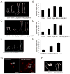
Figure 3
Figure 3. HMR acts specifically in phytochrome signaling pathways between PHY and DET1
(A) Images of 4-day old Col-0, hmr-2, phyB-9, hmr-2/phyB-9 double mutant seedlings grown under 8 μmol m-2 sec-1 R light. (B) Hypocotyl length measurements of seedlings in (A). (C) Images of 4-day old Col-0, hmr-2, phyA-211, hmr-2/phyA-211 double mutant seedlings grown under 3.6 μmol m-2 sec-1 FR light. (D) Quantitative hypocotyl length measurements of seedlings in (C). (E) Images of 4-day old dark-grown PHYBYH, hmr-1/PHYBYH, and Ler seedlings. (F) Hypocotyl measurements of 4-day old dark-grown PHYBYH, hmr-1/PHYBYH, and Ler seedlings. (G) Confocal images of PHYBYH subnuclear localization patterns in 4-old dark-grown PHYBYH and hmr-1/PHYBYH seedlings. DIC and merge images show the location of the nucleus. NB, nuclear body; P, plastid; N, nucleus. (H) Images of 4-day old det1-1 and hmr-1/det1-1 seedlings grown in the dark. Error bars represent standard error. See also Figure S2.
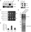
Figure 4
Figure 4. Spatial and temporal expression of HMR RNA and protein
(A) Predicted domain structure of HMR, including a glutamate (GLU) rich region, two bipartite nuclear localization signals (NLSa and NLSb), and a PEST domain. (B) Steady-state HMR mRNA levels in 3-day old Col-0 seedlings grown under D, R, FR, B, and WL measured by qRT-PCR. Error bars represent standard deviation. (C-E) GUS staining of 2-day or 4-day old transgenic lines carrying the HMRp∷GUS construct. The seedlings were grown in the dark (B), red light (C), or far-red light (D). (F) Western blot using anti-HMR antibodies showing HMR proteins in 3-day old seedlings grown under D, R, FR, B, and WL. Levels of RPN6 were used as loading controls.
Figure 5
Figure 5. HMR is required for light-dependent PHYA, PIF1, and PIF3 proteolysis and partially rescues the yeast rad23Δrpn10Δ mutant
(A) Western blot showing PHYA protein levels in 4-day old R light grown Col-0, hmr-2, PBG, hmr-1, and phyB-9. (B) Western blots showing PIF1 and PIF3 protein levels in 4-day R light grown Col-0, hmr-2, PBG, and hmr-1 seedlings. Tubulin was used as a loading control. (C) A growth assay showing serial dilutions of rad23Δrpn10Δ, RAD23rpn10Δ, and HMRrpn10Δ grown in 30°C either with Gal (Galactose) in the upper panel or with Glc (Glucose). The growth defect of rad23Δrpn10Δ was partially rescued only in the presence of Gal, which induces HMR expression in yeast. (D) Western blot showing multiubiquitylated proteins detected by anti-ubiquitin (anti-ubi) antibodies. The SDS-PAGE gel (lower panel) was used as a loading control. (E) UV survival assay using rad23Δ (rad23Δrpn10Δ), RAD23 (RAD23rpn10Δ), and HMR (HMRrpn10Δ). Error bars represent standard error from three independent replica. See also Figure S3.
Figure 6
Figure 6. HMR is localized to both the nucleus and chloroplasts
(A) HMR protein is enriched in both the nuclear and chloroplast protein fractions. Protein extracts of whole plant (T), nuclear (N), or chloroplast (C) fractions from either 2-day old Col-0 seedlings or Broccoli flower buds were separated by SDS-PAGE, and HMR protein was detected by the anti-HMR antibody. Ferrodoxin: Sulfite reductase (SiR) and RNA Pol II were used as controls for the chloroplast and nuclear fractions respectively. (B) Confocal images showing the subcellular localization of HMR in 2-day old R light grown PBG seedlings by immunofluorescent labeling. PHYB∷GFP (green) remains intact under the fixation condition. Both the nuclei (marked by white arrows) and plastid chromosome (marked by yellow arrows) were labeled by DAPI. HMR (red), labeled by anti-HMR antibodies and Alexa 555 conjugated anti-Rabbit secondary antibodies, was detected both in the plastids and the nuclei. (C) Confocal images showing SiR subcellular localization using immunofluorescent labeling. (D) Confocal image showing subnuclear localization of HMR. HMR (red) localizes to foci within the nucleolus and the nucleoplasm. The HMR foci are often adjacent to or sometimes partially overlapping with the PHYB∷GFP (green) NBs. (E) Immunofluorescent labeling using pre-immune serum (red) for anti-HMR antibodies. See also Figure S4.
Figure 7
Figure 7
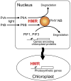
Schematic illustration of a model for HMR functions in the nucleus and chloroplasts. In the nucleus, HMR is essential for PHY NB formation, which is required for the proteolysis of PHYA, PIF1 and PIF3. By controlling PIF1 and PIF3 stability, HMR could indirectly regulate the expression of PIF1/PIF3-controlled genes encoding chloroplast proteins. In chloroplasts, HMR/pTAC12 directly regulates the expression of photosynthetic genes as a transcriptionally active chromosome protein. See also Figure S5.

Similar articles

Cited by

References

    1. Al-Sady B, Kikis EA, Monte E, Quail PH. Mechanistic duality of transcription factor function in phytochrome signaling. Proc Natl Acad Sci (United States of America) 2008;105:2232–2237. - PMC - PubMed
    1. Al-Sady B, Ni W, Kircher S, Schafer E, Quail PH. Photoactivated phytochrome induces rapid PIF3 phosphorylation prior to proteasome-mediated degradation. Mol Cell. 2006;23:439–446. - PubMed
    1. Alonso JM, Stepanova AN, Leisse TJ, Kim CJ, Chen H, Shinn P, Stevenson DK, Zimmerman J, Barajas P, Cheuk R, et al. Genome-wide insertional mutagenesis of Arabidopsis thaliana. Science. 2003;301:653–657. - PubMed
    1. Ang LH, Deng XW. Regulatory hierarchy of photomorphogenic loci: allele-specific and light-dependent interaction between the HY5 and COP1 loci. Plant Cell. 1994;6:613–628. - PMC - PubMed
    1. Bauer D, Viczian A, Kircher S, Nobis T, Nitschke R, Kunkel T, Panigrahi KC, Adam E, Fejes E, Schafer E, et al. Constitutive photomorphogenesis 1 and multiple photoreceptors control degradation of phytochrome interacting factor 3, a transcription factor required for light signaling in Arabidopsis. Plant Cell. 2004;16:1433–1445. - PMC - PubMed

Publication types

MeSH terms