. 2010 Jul 9;142(1):39-51.
doi: 10.1016/j.cell.2010.06.018.

Discovery of a proneurogenic, neuroprotective chemical

Affiliations

Discovery of a proneurogenic, neuroprotective chemical

Andrew A Pieper et al. Cell. .

Abstract

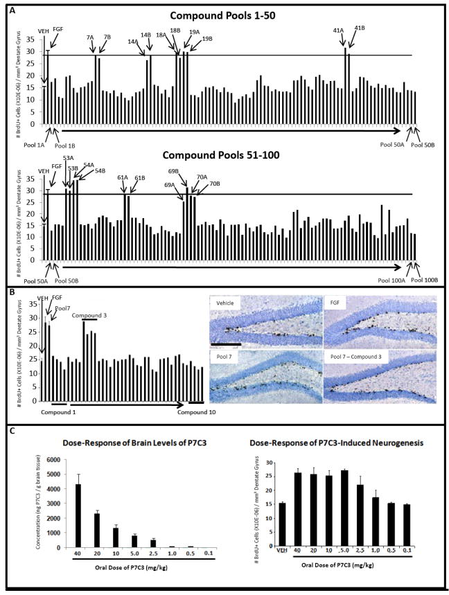
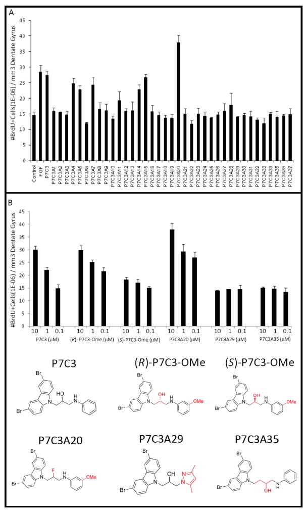
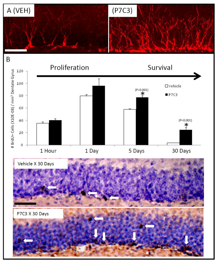
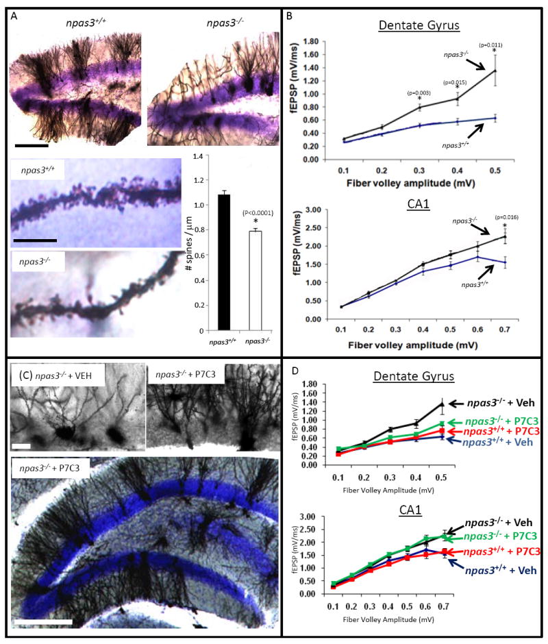
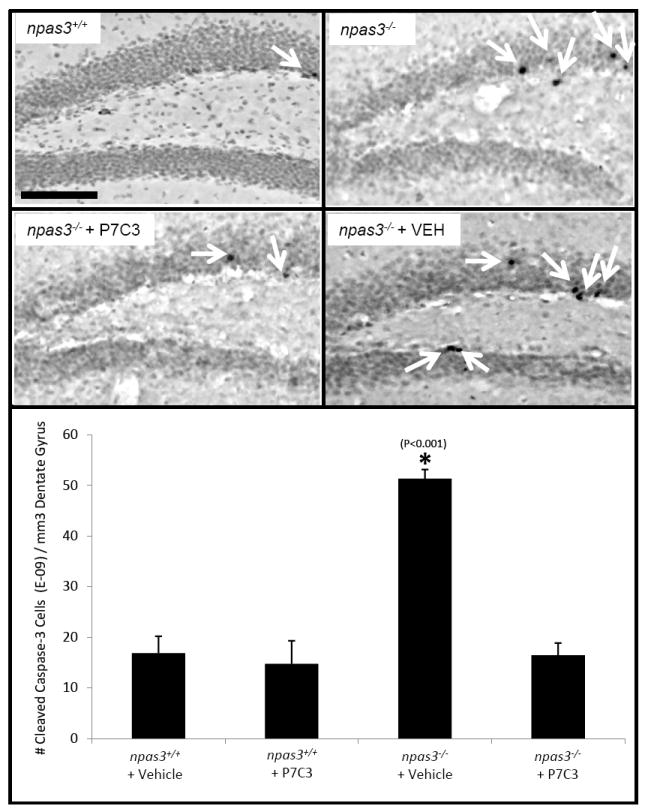
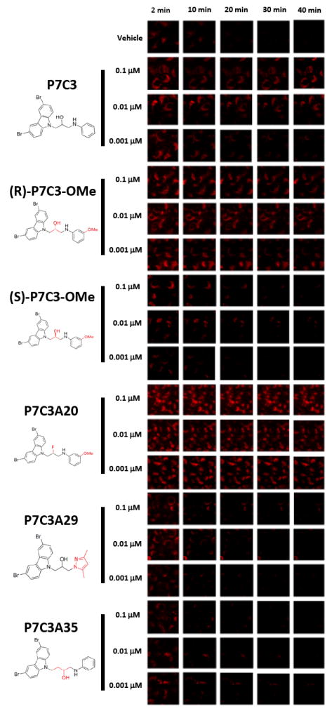
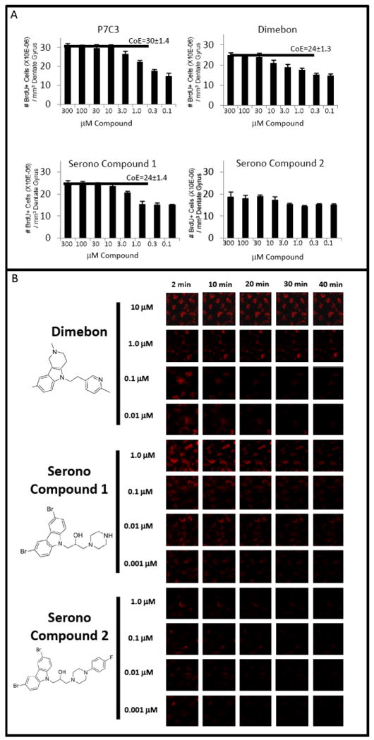
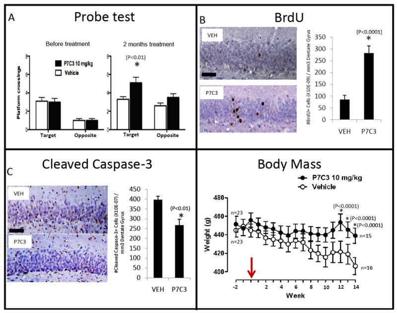
An in vivo screen was performed in search of chemicals capable of enhancing neuron formation in the hippocampus of adult mice. Eight of 1000 small molecules tested enhanced neuron formation in the subgranular zone of the dentate gyrus. Among these was an aminopropyl carbazole, designated P7C3, endowed with favorable pharmacological properties. In vivo studies gave evidence that P7C3 exerts its proneurogenic activity by protecting newborn neurons from apoptosis. Mice missing the gene encoding neuronal PAS domain protein 3 (NPAS3) are devoid of hippocampal neurogenesis and display malformation and electrophysiological dysfunction of the dentate gyrus. Prolonged administration of P7C3 to npas3(-/-) mice corrected these deficits by normalizing levels of apoptosis of newborn hippocampal neurons. Prolonged administration of P7C3 to aged rats also enhanced neurogenesis in the dentate gyrus, impeded neuron death, and preserved cognitive capacity as a function of terminal aging. PAPERCLIP:


Figures

Figure 1
Figure 1. In vivo screen for pro-neurogenic molecules. (See also Figure S1 and Table S1.)
(A) The total number of BrdU+ cells in the SGZ doubled following seven day infusion with FGF-2 relative to vehicle. Each pool of ten compounds was tested for pro-neurogenic efficacy in two mice, and 10 pools displayed efficacy comparable to FGF-2 infusion. The majority of pools displayed no effect. (B) In vivo evaluation in 4 mice each of the ten individual compounds in pool #7 revealed exclusive activity for compound #3. Immunohistochemically-visualized BrdU incorporation in the SGZ is notably greater in animals infused with either pool #7 or compound #3 from pool #7 relative to vehicle-infused animals. All micrographs were taken at the same magnification. (scale bar = 200 μm). (C) P7C3 concentration in mouse brain tissue correlated with oral dosing. Pro-neurogenic efficacy of P7C3 was roughly double that of vehicle at doses ranging from 5 to 40 mg/kg. At decreasing dosage of P7C3 the amount of neurogenesis decreased accordingly, until reaching levels no greater than vehicle at compound doses below 1.0 mg/kg. In all graphs data are expressed as mean +/- SEM.
Figure 2
Figure 2. SAR analysis of structural variants of P7C3
(A) An in vivo SAR study with 37 analogs of P7C3 showed that some analogs had pro-neurogenic activity comparable to the parent compound, whereas others had either no activity or activity intermediate between vehicle and FGF controls. A single compound (P7C3A20) showed enhanced efficacy. (B) Comparison of pro-neurogenic efficacy of analogs of P7C3 showed that the (R)-enantiomer of P7C3-OMe was active, whereas the (S)-enantiomer was not. P7C3A20 exhibited enhanced activity, while P7C3A29 and P7C3A35 suffered structural changes that abolished activity. In all graphs data are expressed as mean +/- SEM.
Figure 3
Figure 3. P7C3 treatment enhances production and survival of neural precursor cells destined to a neuronal fate. (See also Figure S2)
(A) Immunohistochemical staining for DCX was increased in newborn neurons in mice that were administered P7C3. (B) P7C3 enhances hippocampal neurogenesis by promoting survival of newborn neurons without affecting proliferation. In both (A) and (B) both sets of micrographs were taken at the same magnification (scale bar = 50 μm), and are representative of 10 sections from each of 5 mice in each group. Data are expressed as mean +/- SEM.
Figure 4
Figure 4. P7C3 corrects morphological and electrophysiological deficits in npas3-/- dentate gyrus. (See also Figure S3)
(A) Golgi-Cox staining of the dentate gyrus revealed aberrant dendritic arborization in npas3-/- mice relative to wild type littermates (10 sections from each of 5 mice in each group). In addition to reduced dendritic length and branching, npas3-/- dentate gyrus granular neurons also exhibited significantly reduced spine density relative to wild type littermates (*, P < 0.00001, Student’s t test). The top lower power micrographs of dentate gyrus were taken at the same magnification (scale bar = 200 μm). For the bottom two higher power micrographs of dendritic spines the scale bar = 10 μM. (B) In hippocampal slice preparation from npas3-/- mice, synaptic transmission, as assessed by whole field recordings of fEPSPs, was increased both in the outer molecular layer of the dentate gyrus and the CA1 region of the hippocampus relative to wild type. (C) Golgi-Cox staining of dentate gyrus granular neurons showed that prolonged daily treatment of npas3-/- mice with P7C3 enhanced dendritic arborization. Results shown are representative of 10 sections from each of 5 mice in each group. Hi-power micrographs are shown on top (scale bar = 25 μm), and a lower power micrograph illustrating the entire granular layer of the dentate gyrus is shown below (scale bar = 200 μm). (D) Prolonged administration of P7C3 normalized whole field recordings of fEPSPs in the dentate gyrus but not the CA1 region of npas3-/- mice. In all graphs data are expressed as mean +/- SEM.
Figure 5
Figure 5. P7C3 normalizes elevated levels of hippocampal apoptosis in npas3-/- mice
Immunohistochemical detection of CCSP3 showed elevated apoptosis in the dentate gyrus of npas3-/- deficient animals (*, P<0.001). This was normalized by prolonged treatment with P7C3. 3 adult animals were tested in each group. Data are expressed as mean +/- SEM.
Figure 6
Figure 6. P7C3 and its analogs preserve mitochondrial membrane potential in parallel to pro-neurogenic activity (see also Figure S4)
U2OS cells were loaded with tetramethylrhodamine methyl ester (TMRM) dye and then exposed to the calcium ionophore A23187 either in the presence or absence of test compounds. P7C3 preserved mitochondrial membrane potential following exposure to the calcium ionophore A23187 in a dose dependent manner. The protective effect of P7C3 was enantiomeric specific. The (R)-enantiomer of P7C3-OMe blocked dye release at levels as lowas 1nM, whereas the (S)-enantiomer failed to block dye release even at the highest drug dose tested (100nM). The more active, pro-neurogenic analog (P7C3A20) exhibited dye release protection at all doses tested, yet analogs devoid of pro-neurogenic activity (P7C3A29 and P7C3A35) failed to preserve mitochondrial membrane potential at any test dose. Each compound was evaluated in triplicate with similar results.
Figure 7
Figure 7. Comparison of P7C3 to other putative anti-apoptotic agents
(A) Adult mice (n=4 for each group) were administered equivalent dose escalations of P7C3, Dimebon and Serono Compounds 1 and 2 for one week. Dimebon was observed to enhance hippocampal neurogenesis, yet with diminished potency and efficacy relative to P7C3. Serono Compound 1, which was more active than Serono Compound 2 in inhibiting BID-mediated cytochrome c release from isolated mitochondria (Bombrun et al., 2003), was similarly superior to Serono Compound 2 in enhancing in vivo neurogenesis. The pro-neurogenic efficacy of Serono Compound 1 was comparable to Dimebon and diminished relative to P7C3. Horizontal bars designate ceiling of efficacy (CoE) levels for each compound. There is no CoE determination for Soreno Compound 2 because this agent did not significantly enhance neurogenesis. Data are expressed as mean +/- SEM. (B) Cultured U2OS cells were loaded with TMRM dye and exposed to a calcium ionophore in the presence of different doses of Dimebon, Serono Compound 1 or Serono Compound 2. Dimebon protected the mitochondrial membrane potential of U2OS cells only at the relatively high doses of 10 and 1uM. The more active, pro-neurogenic Serono Compound 1 showed an enhanced ability to preserve mitochondrial membrane potential relative to the less active Serono Compound 2. Serono Compound 1 lost activity between 10 and 1nM doses.
Figure 8
Figure 8. P7C3 enhances hippocampal neurogenesis, ameliorates cognitive decline, and prevents weight loss in terminally aged rats. (See also Figure S5.)
(A) Prior to treatment, both groups (n=23 for each group) showed similar frequency of crossings through the goal platform. After 2 months of treatment, however, P7C3-treated rats displayed a statistically significant increase of crossings through the goal platform area relative to vehicle-treated rats. (B) P7C3-treated rats displayed significantly enhanced hippocampal neurogenesis, as assessed by BrdU incorporation, relative to vehicle-treated rats. Many more of the BrdU-labeled cells were noted to have migrated into the granular layer in P7C3-treated rats in comparison to vehicle-treated animals, consistent with their functional incorporation into the dentate gyrus as properly wired neurons. (C) Relative to vehicle-treated animals, P7C3-treated rats displayed a significantly lower number of cleaved caspase 3-positive cells in the dentate gyrus, indicating that P7C3 was capable of inhibiting apoptosis in the aged rat brain. Data are expressed as mean +/- SEM. (D) Relative to vehicle-treated animals, P7C3-treated rats were observed to maintain stable body weight as a function of terminal aging. In all graphs data are expressed as mean +/- SEM.

Similar articles

Cited by

References

    1. Altman J. Are new neurons formed in the brains of adult mammals? Science. 1962;135:1127–1128. - PubMed
    1. Altman J, Das GD. Autoradiographic and histological evidence of postnatal hippocampal neurogenesis in rats. J Comp Neurol. 1965;124:319–335. - PubMed
    1. Bachurin S, Bukatina E, Lermontova N, Tkachenko S, Afanasiev A, Grigoriev V, Grigorieva I, Ivanov Y, Sablin S, Zefirov N. Antihistamine agent Dimebon as a novel neuroprotector and a cognition enhancer. Ann N Y Acad Sci. 2001;939:425–435. - PubMed
    1. Bachurin SO, Shevtosa EP, Kireeva EG, Oxenkrug GF, Sablin SO. Mitochondria as a target for neurotoxins and neuroprotective agents. Ann N Y Acad Sci. 2003;993:334–344. - PubMed
    1. Boldrini M, Underwood MD, Hen R, Rosoklija GB, Dwork AJ, John Mann J, Arango V. Antidepressants increase neural progenitor cells in the human hippocampus. Neuropsychopharmacology. 2009;34:2376–2389. - PMC - PubMed

Publication types

MeSH terms