. 2010 Jul 9;142(1):65-76.
doi: 10.1016/j.cell.2010.06.021.

Identification of KIAA1018/FAN1, a DNA repair nuclease recruited to DNA damage by monoubiquitinated FANCD2

Affiliations

Identification of KIAA1018/FAN1, a DNA repair nuclease recruited to DNA damage by monoubiquitinated FANCD2

Craig MacKay et al. Cell. .

Abstract

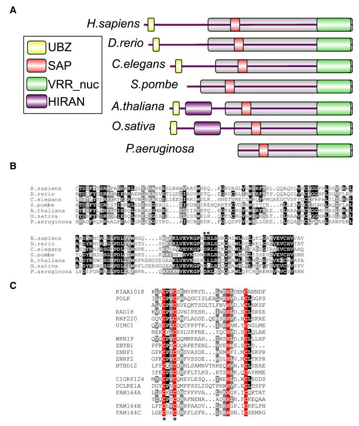
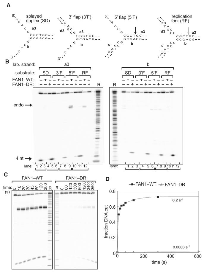
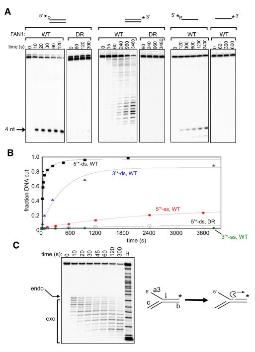
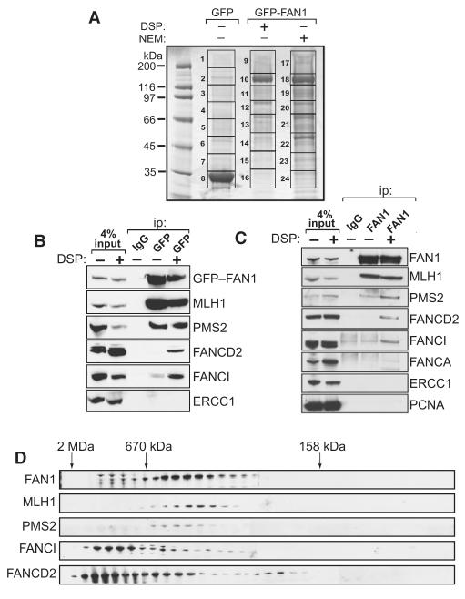
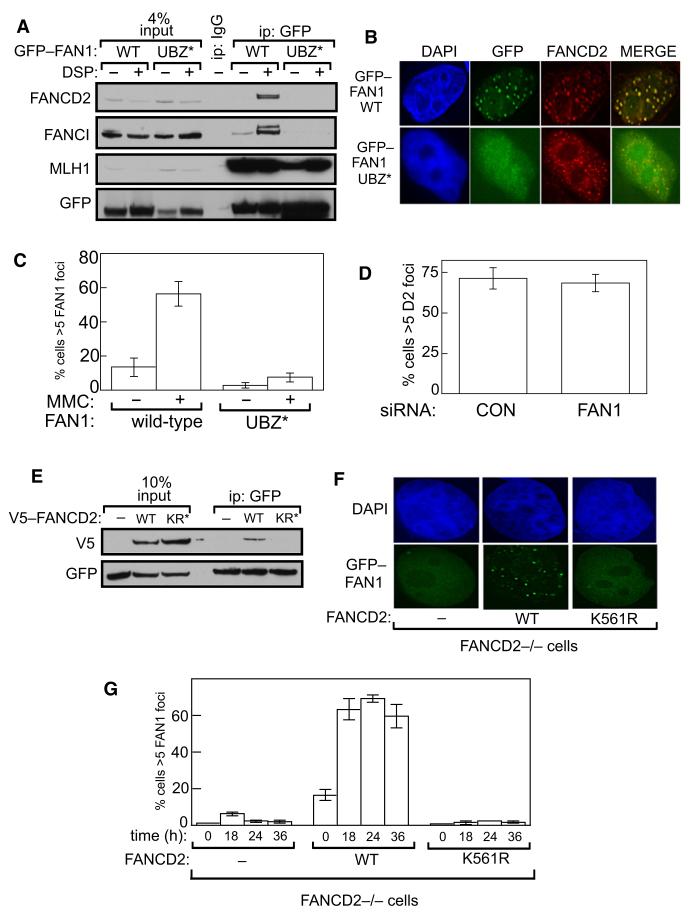
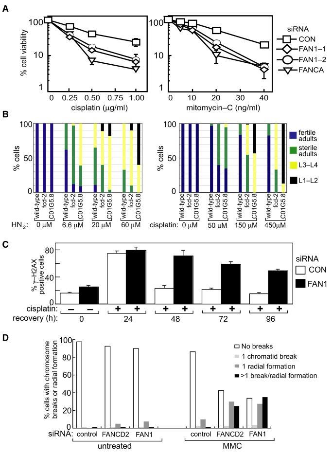
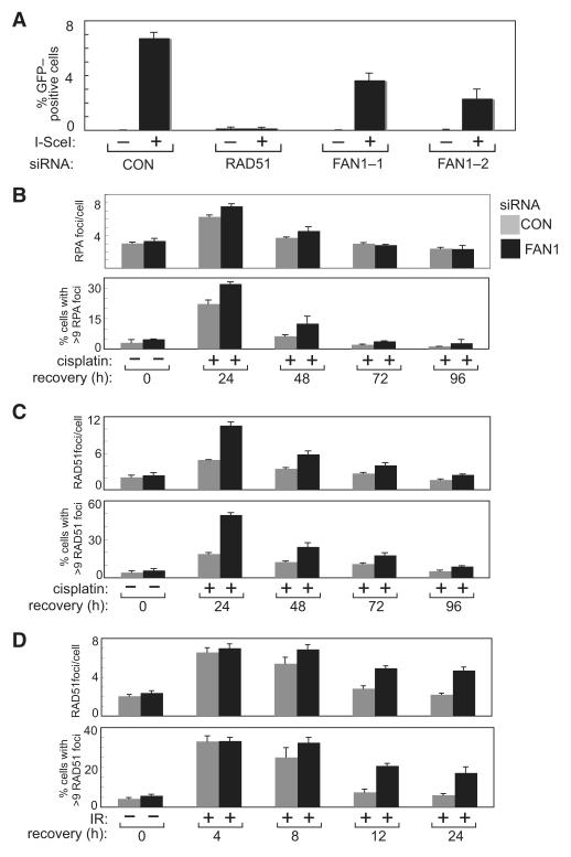
DNA interstrand crosslinks (ICLs) are highly toxic because they block the progression of replisomes. The Fanconi Anemia (FA) proteins, encoded by genes that are mutated in FA, are important for repair of ICLs. The FA core complex catalyzes the monoubiquitination of FANCD2, and this event is essential for several steps of ICL repair. However, how monoubiquitination of FANCD2 promotes ICL repair at the molecular level is unknown. Here, we describe a highly conserved protein, KIAA1018/MTMR15/FAN1, that interacts with, and is recruited to sites of DNA damage by, the monoubiquitinated form of FANCD2. FAN1 exhibits endonuclease activity toward 5' flaps and has 5' exonuclease activity, and these activities are mediated by an ancient VRR_nuc domain. Depletion of FAN1 from human cells causes hypersensitivity to ICLs, defects in ICL repair, and genome instability. These data at least partly explain how ubiquitination of FANCD2 promotes DNA repair.


Figures

Figure 1
Figure 1. The KIAA1018/MTMR15/FAN1 Family of Proteins
(A) Schematic representation of the domain architecture of KIAA1018/MTMR15/FAN1 orthologs from different species. The relevant protein identification codes are as follows: Homo sapiens Q9Y2M0, Danio rerio Q1LWH4, Caenorhabditis elegans P90740, Schizosaccharomyces pombe Q9Y804, Arabidopsis thaliana Q9SX69, Oryza sativa B9FRR6, and Pseudomonas aeruginosa Q9I2N0. (B) Alignment of the VRR_nuc domain of FAN1. Identical residues are shaded in black, and similar residues are shaded in gray. The asterisks denote conserved residues Asp981 and Arg982 mutated in the FAN1-DR mutant. (C) Alignment of the UBZ domain of FAN1. Identical residues are shaded in black, and similar residues are shaded in gray. The conserved Cys and His residues that define the two dyads of the UBZ domain are shaded in red. Asterisks denote the conserved Cys44 and Cys47 residues in the first dyad.
Figure 2
Figure 2. FAN1 Has Structure-Specific Endonuclease Activity
Recombinant human FAN1 was incubated with synthetic DNA structures: splayed duplex (SD; oligos a3, b), 3′ flap (3′F; oligonucleotides a3, b, d3), 5′ flap (5′F; oligos a3, b, c), or a replication fork (RF)-like structure (oligos a3, b, c, d3), each radioactively 5′ 32P-labeled on the strands indicated. WT refers to wild-type FAN1, and DR refers to the Asp981Ala-Arg982Ala FAN1 mutant. (A) Schematic diagram of the DNA substrates used in (B). Sites of DNA cleavage are indicated by arrows. (B) Reaction products (10 min incubation) were subjected to denaturing PAGE. Purine-specific chemical sequencing ladders (R) were derived from oligonucleotides a3 or b. (C) FAN1 was incubated with the 5′ flap shown in (A) for the time indicated (s, seconds), and reaction products were subjected to denaturing PAGE. (D) Progress curves of cleavage of the 5′ flap construct incubated with wild-type (black squares) and DR (gray triangles) mutant FAN1. The data have been fitted to a single (DR) or double (wild-type) exponential functions (lines). From these data, we have calculated observed rates of cleavage of >0.2 s−1 and 0.0003 s−1 for wild-type and DR enzymes, respectively. See also Figure S2.
Figure 3
Figure 3. FAN1 has 5′ Exonuclease Activity
Recombinant human FAN1 was incubated for the time indicated (s, seconds) with dsDNA (oligonucleotide a3, a3-cp), ssDNA (oligonucleotide a3), or a 5′ flap (5′F; oligonucleotides a3, b, c) radioactively 5′ or 3′ 32P-labeled on the a3 strand as shown (asterisks). WT refers to wild-type FAN1, and DR refers to the Asp981Ala-Arg982Ala FAN1 mutant. Reaction products were subjected to denaturing PAGE. (A) Cleavage of linear DNA substrates. (B) The cleavage products were quantitated. “Fraction DNA cut” is the ratio of the relevant cleavage product to total DNA (cleaved plus uncleaved DNA). The data are plotted as a function of time and are fitted to single or double exponential functions. (C) Activity of WT FAN1 on radioactively 3′ 32P-labeled 5′ flap. R refers to a purine-specific chemical sequencing ladder derived from the labeled strand. See also Figure S3.
Figure 4
Figure 4. FAN1 Interacts with DNA Repair Proteins
(A) HEK293 Flp-In cells that stably express GFP-FAN1 were lysed in the presence of dithiobis (succinimidyl propionate) (DSP) or N-ethyl maleimide (NEM). These extracts together with extracts of cells that express GFP only were subjected to immunoprecipitation with GFP-Trap beads, and after extensive washing precipitates were subjected to SDS-PAGE. The gel was fixed and stained with Colloidal Blue. The gel lanes were cut into slices, as indicated, and the proteins were digested with trypsin before mass spectrometric fingerprinting. (B) HEK293 Flp-In cells that stably express GFP-FAN1 were lysed in the presence or absence of DSP and extracts were subjected to immunoprecipitation with control anti-HA (IgG) or GFP-Trap beads. Precipitates were analyzed by western blotting with the antibodies indicated. Input represents 4% of the extract used for immunoprecipitation. (C) HEK293 cells were lysed in the presence or absence of DSP, and extracts were subjected to immunoprecipitation with anti-HA (IgG) or with anti-FAN1 antibodies. Precipitates were analyzed by western blotting with the antibodies indicated. Input represents 4% of the extract used for immunoprecipitation. (D) Extracts of HEK293 cells were analyzed by size-exclusion chromatography on a HiLoad 26/60 Superdex 200 column in buffer containing 0.2 M NaCl, and every third fraction was denatured and analyzed by western blotting with the indicated antibodies. The elution positions of Dextran blue (2 MDa), thyroglobulin (670 kDa), and bovine γ-globulin (158 kDa) are shown. See also Table S1.
Figure 5
Figure 5. The UBZ Domain of FAN1 Binds Monoubiquitinated FANCD2
(A) HEK293 cells were transiently transfected with pcDNA5.1-GFP-FAN1 wild-type (WT) or pcDNA5.1-GFP-FAN1-UBZ* (Cys44A/Cys47A). After 48 hr, cells were lysed in the presence or absence of DSP, and anti-GFP precipitates were analyzed by western blotting with the antibodies indicated. “Input” represents cell extracts. (B) U2OS cells, grown on glass coverslips, were transiently transfected with pcDNA5.1-GFP, pcDNA5.1-GFP-FAN1 wild-type (WT), or pcDNA5.1-GFP-FAN1-UBZ* (Cys44A/Cys47A). Cells were treated, or not, with MMC, and after 16 hr GFP-FAN1 foci and FANCD2 foci were detected. (C) Quantitation of data from (B). The number of cells with more than five GFP-FAN1 foci in a sample of 500 cells was counted. (D) U2OS cells were transfected with control siRNA or siRNA targeting FAN1, and the number of cells with more than five FANCD2 foci was quantitated after exposure of cells to MMC. (E) HEK293 cells stably expressing GFP-FAN1 were transiently transfected with pDEST40-lacZ (“–”), pDEST40-V5-FANCD2 wild-type (WT), or pDEST40-V5-FANCD2 K561R. After 48 hr, cells were lysed in the presence of DSP, and anti-GFP precipitates were analyzed by western blotting with the antibodies indicated. (F) FANCD2−/− (PD20) cells stably transfected with empty vector (–), FANCD2 wild-type (WT), or FANCD2 K561R were transiently transfected with GFP-FAN1. Cells were treated with MMC for 18 hr and then fixed, and GFP-FAN1 foci were visualized. (G) Same as (F), except that cells were treated with MMC for the times indicated, and the number of cells with more than five FAN1 foci were quantitated after exposure of cells to MMC. Data in (C), (D), and (G) are represented as mean ± SEM. See also Figure S4.
Figure 6
Figure 6. FAN1 Is Required for ICL Repair
(A) HEK293 cells were transfected with the siRNAs indicated. Clonogenic survival assays were carried out with cisplatin or mitomycin-C (see the Experimental Procedures). For each siRNA, cell viability of untreated cells is defined as 100%. (B) Synchronized L1 larva stage animals of the relevant genotype were incubated with the indicated concentrations of nitrogen mustard (HN2) or cisplatin. After 48 hr, the extent of developmental progression of the worms was scored, by counting the number of worms in the adult and various larval stages (L1–L2, L3–L4). Adult worms were scored as fertile if they contained fertilized eggs and as sterile if they did not. (C) HEK293 cells transfected with control siRNA or FAN1 siRNA (FAN1-1) were treated with cisplatin (1 μg/ml) for 2 hr and then allowed to recover for the times indicated. The proportion of cells in each population with more than two γ-H2AX foci at each time point (“γ-H2AX positive”) was determined. The experiment was done three times, and a representative experiment is shown. (D) The frequency of chromosome breaks and radial chromosomes in metaphase spreads of HEK293 cells transfected with control siRNA or FAN1-1 siRNA was measured before and after exposure to MMC (25 ng/ml; 18 hr) was measured as described previously (Deans and West, 2009). Data in (A) and (C) are represented as mean ± SEM. See also Figure S5.
Figure 7
Figure 7. Effect of FAN1 Depletion on HR and Focus Formation by RAD51 and RPA
(A) U2OS cells, in which an 18 bp sequence recognized by I-SceI was placed between tandem mutant copies of the gene encoding GFP, were transfected with control siRNA (luciferase) and/or siRNAs specifically targeting FAN1 (FAN1-1 or FAN1-2) or RAD51. After 48 hr, cells were transfected with a plasmid expressing I-SceI or with an empty vector, and 24 hr later cells were tested for GFP expression by FACS analysis. The frequency of HR in cells transfected with the various siRNAs was calculated relative to cells transfected with control siRNA. (B) U2OS cells transfected with control siRNA or FAN1 siRNA (FAN1-1) were treated with cisplatin (1 μg/ml) for 2 hr and then allowed to recover for the times indicated. Cells were then fixed, permeabilized, washed, and blocked before incubation with anti-RPA antibodies. Coverslips were incubated with secondary antibodies, mounted on glass slides, and visualized. The average number of RPA foci per cell was analyzed. The experiment was done three times, and a representative experiment is shown. (C) Same as (B), except that cells were stained with anti-RAD51 antibodies. (D) Same as (C), except that cells were exposed to IR (3 Gy) and then allowed to recover for the times indicated. Data represented as mean ± SEM. See also Figure S6.

Comment in

Similar articles

Cited by

References

    1. Akkari YMN, Bateman RL, Reifsteck CA, Olson SB, Grompe M. DNA replication is required To elicit cellular responses to psoralen-induced DNA interstrand cross-links. Mol. Cell. Biol. 2000;20:8283–8289. - PMC - PubMed
    1. Bergstralh DT, Sekelsky J. Interstrand crosslink repair: can XPF-ERCC1 be let off the hook? Trends Genet. 2008;24:70–76. - PubMed
    1. Bhagwat N, Olsen AL, Wang AT, Hanada K, Stuckert P, Kanaar R, D’Andrea A, Niedernhofer LJ, McHugh PJ. XPF-ERCC1 participates in the Fanconi anemia pathway of cross-link repair. Mol. Cell. Biol. 2009;29:6427–6437. - PMC - PubMed
    1. Bienko M, Green CM, Crosetto N, Rudolf F, Zapart G, Coull B, Kannouche P, Wider G, Peter M, Lehmann AR, et al. Ubiquitin-binding domains in Y-family polymerases regulate translesion synthesis. Science. 2005;310:1821–1824. - PubMed
    1. Brenner S. The genetics of Caenorhabditis elegans. Genetics. 1974;77:71–94. - PMC - PubMed

Publication types

MeSH terms