

Review
doi: 10.1016/j.molcel.2010.06.017.
The PARP side of the nucleus: molecular actions, physiological outcomes, and clinical targets
Affiliations
- PMID: 20603072
- PMCID: PMC2923840
- DOI: 10.1016/j.molcel.2010.06.017
Item in Clipboard
Review
The PARP side of the nucleus: molecular actions, physiological outcomes, and clinical targets
Mol Cell.
.
Abstract
The abundant nuclear enzyme PARP-1, a multifunctional regulator of chromatin structure, transcription, and genomic integrity, plays key roles in a wide variety of processes in the nucleus. Recent studies have begun to connect the molecular functions of PARP-1 to specific physiological and pathological outcomes, many of which can be altered by an expanding array of chemical inhibitors of PARP enzymatic activity.
2010 Elsevier Inc. All rights reserved.
Figures
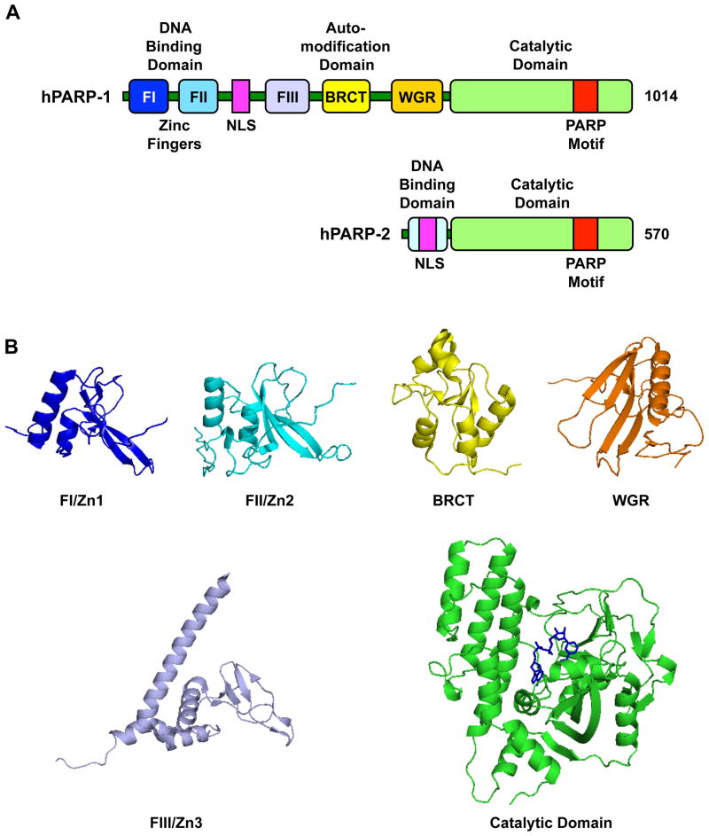
(A) Schematic representation of human PARP-1 and PARP-2 with the functional domains noted in the text. (B) Structures of the six structural and functional domains in human PARP-1. FI (PDB 2DMJ), FII (PDB 2CS2), FIII (PDB 2RIQ), BRCT (PDB 2COK), WGR (PDB: 2CR9), catalytic domain (PDB 1A26; NAD+ has been modeled in based on a structure of diptheria toxin, PDB 1TOX).
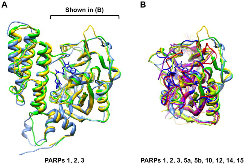
(A) Alignment of the catalytic domain structures from mammalian PARP-1 (PDB 1A26; green), PARP-2 (PDB 1GSO; yellow), and PARP-3 (PDB 3FHB; blue). (B) Alignment of the catalytic domain structures from mammalian PARPs 1, 2, 3, 5a, 5b, 10, 12, 14, 15 (PDB 1A26, 1GSO, 3FHB, 2RF5, 3KR7, 3HKV, 2PQF, 3GOY, 3GEY, respectively). In (A) and (B), NAD+ has been modeled in based on a structure of diptheria toxin (PDB 1TOX).
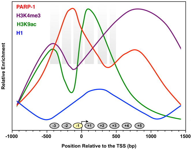
Schematic of average genomic ChIP and nucleosome mapping data across the promoters of the most highly expressed genes (top quartile) in cells. The graphs are based on data from the literature: PARP- 1 and H1 (Krishnakumar et al., 2008), H3K4me3 (Barski et al., 2007), H3K9ac (Wang et al., 2008b), and nucleosome positioning (Schones et al., 2008).
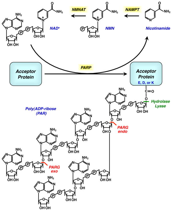
Chemical structures of NAD+, PAR, and metabolites. The enzymes that catalyze the synthesis of NAD+ in the mammalian salvage pathway are shown. The enzymatic actions of PARP, PARG, (ADP-ribosyl) protein hydrolase, and (ADP-ribosyl) protein lyase are also indicated.
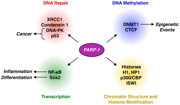
PARP-1 interacts with and PARylates proteins involved in DNA repair, transcription, DNA methylation, and the regulation of chromatin structure and histone modification to control physiological and pathological outcomes.
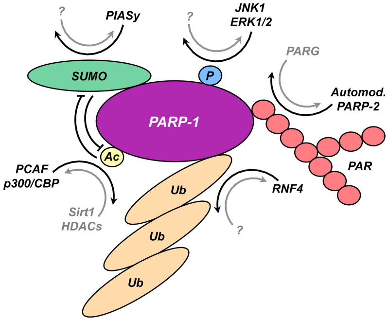
Schematic representation of PARP-1 modifications: PARylation, phosphorylation, acetylation, SUMOylation, ubiquitylation, as described in the text.
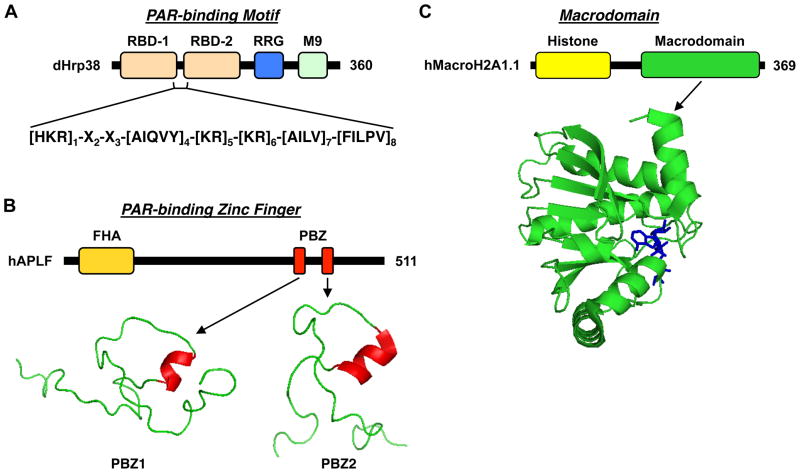
(A) PAR binding motifs, as found in dHrp38, a protein that regulates alternative splicing of RNA transcripts. (B) PAR-binding zinc fingers (PBZs), as found in hAPLF, a protein involved in DNA damage checkpoints. The structures of the two PBZs from hAPLF are shown. (C) Macrodomains, as found in macroH2A1.1, a histone variant involved in setting the chromatin environment. The structure of the macrodomain of macroH2A1.1 bound to ADP-ribose is shown.
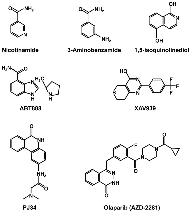
Structures of PARP inhibitors.
References
-
- Aguilar-Quesada R, Munoz-Gamez JA, Martin-Oliva D, Peralta A, Valenzuela MT, Matinez-Romero R, Quiles-Perez R, Menissier-de Murcia J, de Murcia G, Ruiz de Almodovar M, Oliver FJ. Interaction between ATM and PARP-1 in response to DNA damage and sensitization of ATM deficient cells through PARP inhibition. BMC Mol Biol. 2007;8:29. - PMC - PubMed
-
- Ahel I, Ahel D, Matsusaka T, Clark AJ, Pines J, Boulton SJ, West SC. Poly(ADP-ribose)-binding zinc finger motifs in DNA repair/checkpoint proteins. Nature. 2008;451:81–85. - PubMed
-
- Alvarez-Gonzalez R, Jacobson MK. Characterization of polymers of adenosine diphosphate ribose generated in vitro and in vivo. Biochemistry. 1987;26:3218–3224. - PubMed
Publication types
MeSH terms
Substances
Grants and funding
LinkOut - more resources
Full Text Sources
Other Literature Sources
Miscellaneous
