. 2010 Oct;139(4):1385-96.
doi: 10.1053/j.gastro.2010.06.069. Epub 2010 Aug 7.

FGF receptors 1 and 2 control chemically induced injury and compound detoxification in regenerating livers of mice

Affiliations

FGF receptors 1 and 2 control chemically induced injury and compound detoxification in regenerating livers of mice

Friederike Böhm et al. Gastroenterology. 2010 Oct.

Abstract

Background & aims: Fibroblast growth factor receptor 4 (FGFR4) controls bile acid metabolism and protects the liver from fibrosis, but the roles of FGFR1 and FGFR2 in the adult liver are largely unknown. We investigated the functions and mechanisms of action of these receptors in liver homeostasis, regeneration, and fibrosis.

Methods: We generated mice with hepatocytes that lack FGFR1 and FGFR2 and subjected them to acute and chronic carbon tetrachloride-induced liver injury and partial hepatectomy; mice were also injected with FGF7. We performed histology, histomorphometry, real-time reverse transcription polymerase chain reaction, and immunoblot analyses.

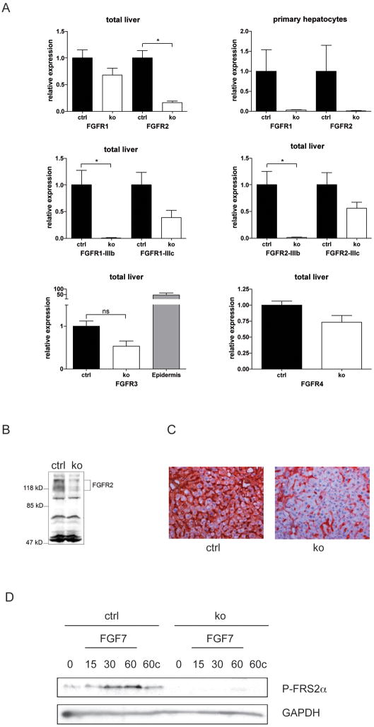
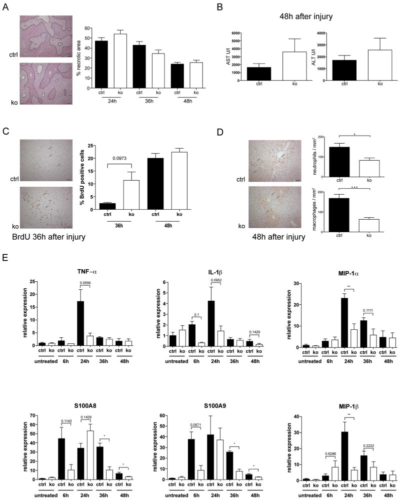
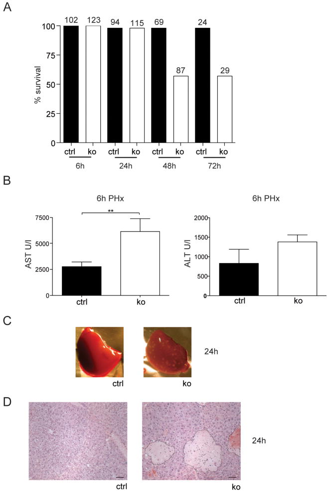
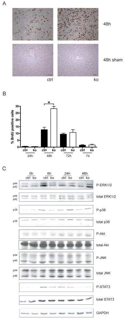
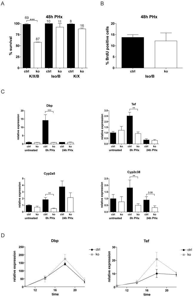
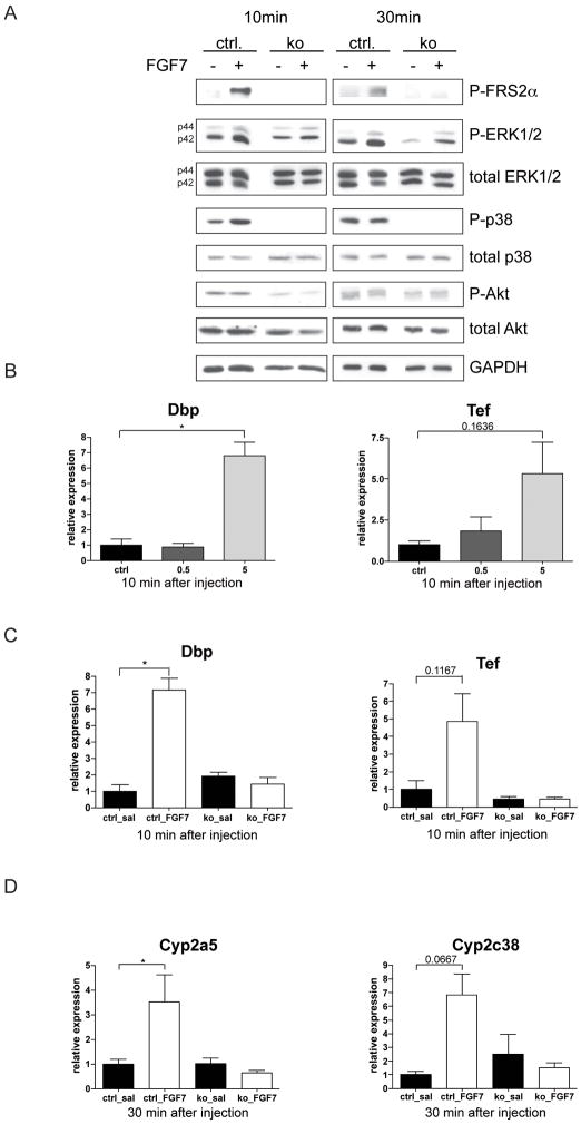
Results: In hepatocytes, loss of FGFR1 and FGFR2 eliminated responsiveness to FGF7 and related FGF family members but did not affect toxin-induced liver injury and fibrosis. However, mortality after partial hepatectomy increased because of severe hepatocyte necrosis. These effects appeared to be mediated by a failure of hepatocytes to induce the expression of the transcriptional regulators Dbp and Tef upon liver surgery; this affected expression of their target genes, which encode detoxifying cytochrome P450 enzymes. We found that Dbp and Tef expression was directly controlled by FGFR signaling in hepatocytes. As a consequence of the reduced expression of genes that control detoxification, the liver tissue that remained after partial hepatectomy failed to efficiently metabolize endogenous compounds and the drugs applied for anesthesia/analgesia.

Conclusions: We identified a new, cytoprotective effect of FGFR1 and FGFR2 in the regenerating liver and suggest the use of recombinant FGF7 to increase survival of patients after surgical resection of large amounts of liver tissue.


Conflict of interest statement

Disclosures: The authors have no conflict of interest

Figures

Fig. 1
Fig. 1. Verification of the loss of FGFR1 and FGFR2 in hepatocytes
(A) RNAs from total liver or primary hepatocytes of Alb-R1/R2 (ko) and control mice (ctrl) were analyzed for Fgfr or Gapdh expression using qRT-PCR. Expression levels in control mice were arbitrarily set as 1. RNA from mouse epidermis was used as a positive control for FGFR3 expression. (B) Lysates from total liver of Alb-R2-IIIb and control mice were analyzed by western blotting for expression of FGFR2. Differently glycosylated forms of FGFR2 are indicated. (C) Sections from the liver of Alb-R2-IIIb and control mice were analyzed by immunohistochemistry for expression of FGFR2. (D) Primary hepatocytes from control and Alb-R1/R2 mice were serum-starved, treated with 10 ng/ml FGF7, harvested at the indicated time points and analyzed by western blotting for phosphorylated FRS2α and GAPDH. Cells incubated for 60 min in the absence of FGF7 were used as control (60c).
Fig. 2
Fig. 2. Loss of FGFR1 and FGFR2 in hepatocytes protects from CCl4-induced inflammation
(A–E) Mice were injected once with CCl4 in mineral oil and sacrificed at different time points. (A) Representative hematoxylin/eosin-stained sections are shown. Necrotic area can be distinguished from normal liver tissue by the brighter colour and the inflammatory cell infiltrates (encircled areas). It was determined by measuring 4–5 independent microscopic fields (N≥5 per genotype) and is indicated as percent of total liver area. Bar: 100μm. (B) AST and ALT levels were determined in the serum 48h after CCl4 injection. N≥5 per genotype. (C) Liver sections from BrdU-injected animals were stained with an antibody against BrdU. Representative sections from Alb-R1/R2 mice and controls 36h after CCl4 injection are shown. The percentage of BrdU-positive cells was determined by counting 4–5 independent microscopic fields. N≥5 mice per time point. Bar: 50μm. (D) Liver sections were stained with antibodies against neutrophils (Ly6G) or macrophages (ER-MP23). Representative pictures are shown. Inflammatory cells were counted in 4–5 independent microscopic fields (200 × magnification, N≥5 per genotype). Bar: 50μm. (E) RNAs from the liver of control and Alb-R1/R2 mice at different time points after CCl4 injection were analyzed by qRT-PCR for the levels of IL-1β, TNF-α, S100A8, S100A9, MIP-1α, or MIP-1β mRNAs. Rps29 was used for normalization.
Fig. 3
Fig. 3. FGFR1 and FGFR2 cooperate to enhance survival of mice and to prevent hepatocyte necrosis after PH
Mice were subjected to PH. (A) The percentage of surviving animals at different time points after PH is indicated. Numbers above the bars indicate the number of animals observed at each time point per genotype. (B) AST and ALT levels in the serum were determined 6h after PH. N≥5 per genotype. (C,D) Macroscopic (C) and histological analysis (D) revealed the presence of necrotic lesions in the liver of Alb-R1/R2 mice 24h after PH (N≥12 per genotype). Bar: 50μm.
Fig. 4
Fig. 4. Delayed activation of major signalling pathways and enhanced proliferation of hepatocytes after PH in surviving Alb-R1/R2 mice
(A) Cell proliferation in the liver at 48h after PH was assessed by BrdU incorporation. Representative sections from injured liver (48h after PH) are shown. Bar: 50μm. (B) The percentage of proliferating cells was determined by counting 4–5 independent microscopic fields/per liver at 200× magnification, N>6 per genotype and time point. (C) Liver lysates from control and Alb-R1/R2 mice before and at different time points after PH were analyzed by western blotting for the levels of total and phosphorylated ERK1/2, p38, Akt, JNK1/2, STAT3 and GAPDH. Each lysate was obtained from pooled livers of three animals per time point and genotype. All results shown on this figure were obtained with the same lysates. Results were reproduced in an independent experiment using lysates from different animals.
Fig. 5
Fig. 5. FGFR1 and FGFR2 signaling is important for compound detoxification in the regenerating liver
(A) Mice were subjected to PH. One group was anaesthetized with ketamine/xylazine followed by buprenorphine treatment (K/X/B), in the second group ketamine/xylazine was replaced by isoflurane (Iso/B), and in the third group buprenorphine was omitted after ketamine/xylazine anaesthesia (K/X). The percentage of surviving animals at different time points after PH is indicated. Numbers above the bars indicate the number of animals observed at each time point per genotype and treatment group. (B) Cell proliferation was assessed in mice of the Iso/B group 48h after PH using BrdU incorporation. N>6 per genotype and treatment group. (C) Expression of Dbp, Tef, Cyp2a5, and Cyp2c38 was analyzed by qRT-PCR using RNAs from livers of untreated Alb-R1/R2 mice and control animals. RNAs from the liver tissue, which was removed upon PH (0h PH), or from the remaining liver 24h after PH were analyzed for comparison. (D) Levels of Dbp and Tef mRNAs were analyzed by qRT-PCR in non-injured Alb-R1/R2 and control mice at different day times. Rps29 was used for normalization.
Fig. 6
Fig. 6. FGF7 activates a cytoprotective response in the liver
(A) Liver lysates were prepared from control and Alb-R1/R2 mice 10 and 30 min after injection of saline or 5 μg FGF7 and analyzed by western blotting for the levels of phosphorylated FRS2α and total and phosphorylated ERK1/2, p38, Akt and GAPDH. Each lysate was obtained from a single mouse. For statistical analysis see Suppl. Fig. S6. (B) Control mice were i.p. injected with 0.5 or 5 μg FGF7 or vehicle, sacrificed 10 min after injection, and RNAs from the liver were analyzed by qRT-PCR for expression of Dbp and Tef. (C,D) Alb-R1/R2 mice and control littermates were injected i.p. with saline or 5 μg FGF7. They were sacrificed 10 or 30 min after injection, and RNAs from the liver were analyzed by qRT-PCR for expression of Dbp and Tef (C) or Cyp2a5 and Cyp2c38 (D). Rps29 was used for normalization.

Similar articles

Cited by

References

    1. Taub R. Liver regeneration: from myth to mechanism. Nat Rev Mol Cell Biol. 2004;5:836–47. - PubMed
    1. Fausto N. Involvement of the innate immune system in liver regeneration and injury. J Hepatol. 2006;45:347–9. - PubMed
    1. Michalopoulos GK. Liver regeneration. J Cell Physiol. 2007;213:286–300. - PMC - PubMed
    1. Diehl AM. Liver regeneration. Front Biosci. 2002;7:e301–14. - PubMed
    1. Friedman SL. Molecular regulation of hepatic fibrosis, an integrated cellular response to tissue injury. J Biol Chem. 2000;275:2247–50. - PubMed

Publication types

Substances