. 2010 Aug 2;207(8):1745-55.
doi: 10.1084/jem.20100257. Epub 2010 Jul 5.

Redundant roles for inflammasome receptors NLRP3 and NLRC4 in host defense against Salmonella

Affiliations

Redundant roles for inflammasome receptors NLRP3 and NLRC4 in host defense against Salmonella

Petr Broz et al. J Exp Med. .

Abstract

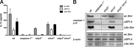
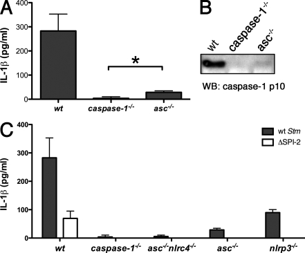
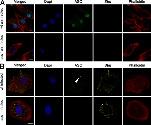
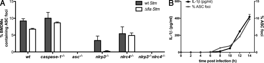
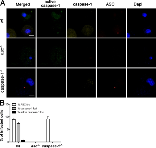
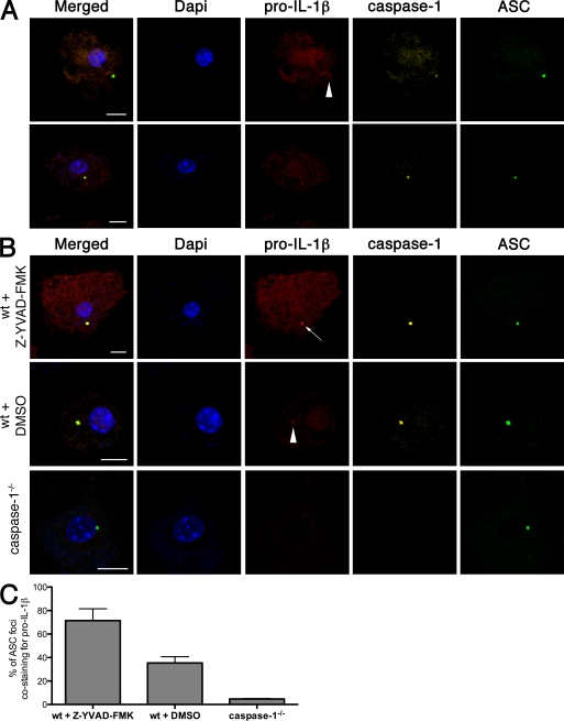
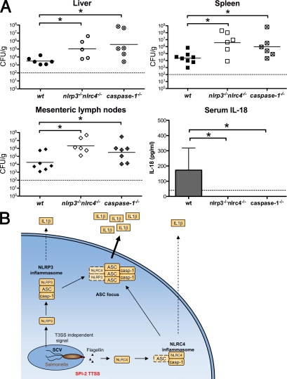
Intracellular pathogens and endogenous danger signals in the cytosol engage NOD-like receptors (NLRs), which assemble inflammasome complexes to activate caspase-1 and promote the release of proinflammatory cytokines IL-1beta and IL-18. However, the NLRs that respond to microbial pathogens in vivo are poorly defined. We show that the NLRs NLRP3 and NLRC4 both activate caspase-1 in response to Salmonella typhimurium. Responding to distinct bacterial triggers, NLRP3 and NLRC4 recruited ASC and caspase-1 into a single cytoplasmic focus, which served as the site of pro-IL-1beta processing. Consistent with an important role for both NLRP3 and NLRC4 in innate immune defense against S. typhimurium, mice lacking both NLRs were markedly more susceptible to infection. These results reveal unexpected redundancy among NLRs in host defense against intracellular pathogens in vivo.


Figures

Figure 1.
Figure 1.
NLRP3 and NLRC4 recognize distinct Stm signals. BM-derived macrophages (BMDMs) of the genotypes indicated were infected with WT Stm for 17 h. Secretion of IL-1β (A) or the processed p10 caspase-1 subunit (B) into the culture supernatant was determined by ELISA and Western blotting, respectively. Equal loading was controlled for by Western blotting for β-actin in the corresponding cell lysates. Gray, white, and black bars in A represent BMDMs infected with WT Stm, a SPI-2 mutant, or a flagellin-deficient strain for 17 h, respectively. Data in A are representative of at least five independent experiments. Data in B are representative of at least two independent experiments. Error bars represent the mean SD of triplicate wells. Black lines indicate that intervening lanes were spliced out.
Figure 2.
Figure 2.
ASC is critical for Stm-induced IL-1β secretion. BMDMs of the genotypes indicated were infected with WT Stm for 17 h. Secretion of IL-1β (A and C) or the processed p10 caspase-1 subunit (B) into the culture supernatant was determined by ELISA and Western blotting, respectively. White bars in C represent BMDMs infected with SPI-2 mutant Stm for 17 h. Equal loading was controlled for by Western blotting for β-actin in the corresponding cell lysates. Data in A and C are representative of five independent experiments. The Western blot in B is representative of three independent experiments. Error bars represent the mean SD of triplicate wells, Statistical significance was determined using the unpaired Mann-Whitney U test. *, P < 0.05.
Figure 3.
Figure 3.
ASC relocalizes in macrophages infected with Stm. Fluorescence microscopy of WT and asc−/− BMDMs not infected (A) or infected with WT Stm for 17 h (B). Cells were stained for ASC, Stm, actin (with phalloidin), and DNA (with DAPI). The arrowhead points to an ASC focus. Images in A and B are representative of three independent experiments (original magnification, 63×). Bars, 10 µm.
Figure 4.
Figure 4.
ASC focus formation requires NLRP3 and NLRC4 and correlates with IL-1β release. (A) Percentage of BMDMs of the genotypes indicated forming ASC foci after infection with WT and Δfla Stm for 17 h. Numbers represent two independent fluorescence microscopy experiments, with at least 500 cells counted in each experiment. (B) Time course of ASC focus formation and IL-1β release in WT BMDMs infected with WT Stm. The percentage of macrophages containing an ASC focus (gray line) was determined by fluorescence microscopy. IL-1β released into the culture supernatant (black line) was determined by ELISA. Data in B are representative of two independent experiments. Error bars represent the mean SD of triplicate wells.
Figure 5.
Figure 5.
Caspase-1 is activated in ASC foci. (A) Fluorescence microscopy of WT, asc−/−, and caspase-1−/− BMDMs infected with WT Stm for 17 h, stained for active caspase-1 (with FAM-YVAD-FMK), caspase-1, ASC, and DNA (with DAPI). Images are representative of three independent experiments. (B) Percentage of infected cells from A containing foci of ASC, caspase-1, and active caspase-1. Quantification represents mean numbers from three independent experiments, with at least 500 cells counted in each experiment. Images in A were acquired at 63× magnification. Cell counts in B were determined at 40× magnification. Error bars represent the mean SD of triplicate experiments. Bars, 10 µm.
Figure 6.
Figure 6.
Pro–IL-1β localizes to ASC foci in a caspase-1–dependent manner. (A) Fluorescence microscopy of WT BMDMs infected with WT Stm for 17 h and then stained for DNA (with DAPI), pro–IL-1β, caspase-1, and ASC. The top row shows a cell with weak pro–IL-1β staining of an ASC focus (arrowhead), whereas the bottom row shows a cell with an ASC- and caspase-1–containing focus that is negative for pro–IL-1β. (B) WT and caspase-1−/− BMDMs infected with WT Stm for 17 h were stained for DNA (with DAPI), pro–IL-1β, caspase-1, and ASC. Where indicated, WT BMDMs were infected in the presence of the caspase-1 inhibitor Z-YVAD-FMK or DMSO (vehicle control). An arrow points to an ASC focus staining strongly for pro–IL-1β. An arrowhead marks an ASC focus with a faint pro-IL-1β signal. (C) Percentage of ASC foci in B costaining for pro–IL-1β. Images and numbers are a representative of three independent experiments with a mean of 50 ASC foci counted in each experiment. Error bars represent the mean SD of triplicate experiments. The antibody used for pro–IL-1β immunofluorescence is a polyclonal antibody recognizing both the uncleaved and mature forms of IL-1β. Images and cell counts were acquired at 63× magnification. Bars, 10 µm.
Figure 7.
Figure 7.
Redundant roles for NLRP3 and NLRC4 in caspase-1 activation during Stm infection in mice. (A) WT, isogenic caspase-1−/−, and nlrp3−/−nlrc4−/− mice were orally infected with 2.4 × 10⁷ CFU of WT Stm. Liver, spleen, and mesenteric lymph nodes were collected at day 5 after infection. Organ homogenates were diluted and plated to determine cfu per gram of tissue. Bars represent the mean bacterial load. Blood was collected at day 4 after infection and serum IL-18 was determined by ELISA. Data represent the mean ± SD of six to eight mice of each genotype. Results are representative of two independent experiments, each done with groups of six to eight mice of each genotype. Statistical significance was determined using the unpaired Mann-Whitney U test. *, P < 0.05. Dashed lines represent the detection limit of the CFU counts or IL-18 ELISA, respectively. (B) The role of the ASC focus in Stm-induced cytokine processing in vitro. Intracellular Stm activate NLRP3 and NLRC4. NLRC4 responds to flagellin injected by the SPI-2 T3SS, whereas NLRP3 responds to an undefined T3SS-independent signal. Both receptors induce the assembly of inflammasomes that can process pro–IL-1β and pro–IL-18 to their mature forms. NLRP3 inflammasome assembly is completely dependent on ASC, whereas NLRC4 may assemble an inflammasome without ASC. We speculate that ASC then promotes inflammasome aggregation into a single subcellular focus. This ASC focus likely mediates the bulk of cytokine processing and might serve to increase cytokine production in response to continuous stimulation of NLRs. SCV, Salmonella-containing vacuole; casp-1, caspase-1.

Similar articles

Cited by

References

    1. Allen I.C., Scull M.A., Moore C.B., Holl E.K., McElvania-TeKippe E., Taxman D.J., Guthrie E.H., Pickles R.J., Ting J.P. 2009. The NLRP3 inflammasome mediates in vivo innate immunity to influenza A virus through recognition of viral RNA. Immunity. 30:556–565 10.1016/j.immuni.2009.02.005 - DOI - PMC - PubMed
    1. Amer A., Franchi L., Kanneganti T.D., Body-Malapel M., Ozören N., Brady G., Meshinchi S., Jagirdar R., Gewirtz A., Akira S., Núñez G. 2006. Regulation of Legionella phagosome maturation and infection through flagellin and host Ipaf. J. Biol. Chem. 281:35217–35223 10.1074/jbc.M604933200 - DOI - PubMed
    1. Beuzón C.R., Méresse S., Unsworth K.E., Ruíz-Albert J., Garvis S., Waterman S.R., Ryder T.A., Boucrot E., Holden D.W. 2000. Salmonella maintains the integrity of its intracellular vacuole through the action of SifA. EMBO J. 19:3235–3249 10.1093/emboj/19.13.3235 - DOI - PMC - PubMed
    1. Brodsky I.E., Monack D. 2009. NLR-mediated control of inflammasome assembly in the host response against bacterial pathogens. Semin. Immunol. 21:199–207 10.1016/j.smim.2009.05.007 - DOI - PubMed
    1. Bryan N.B., Dorfleutner A., Rojanasakul Y., Stehlik C. 2009. Activation of inflammasomes requires intracellular redistribution of the apoptotic speck-like protein containing a caspase recruitment domain. J. Immunol. 182:3173–3182 10.4049/jimmunol.0802367 - DOI - PMC - PubMed

Publication types

MeSH terms

Substances