. 2010 Jul 12;190(1):35-43.
doi: 10.1083/jcb.201004017. Epub 2010 Jul 5.

Long astral microtubules uncouple mitotic spindles from the cytokinetic furrow

Affiliations

Long astral microtubules uncouple mitotic spindles from the cytokinetic furrow

Kathleen E Rankin et al. J Cell Biol. .

Abstract

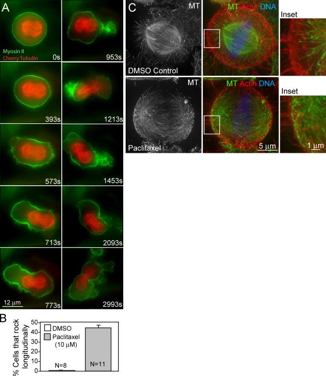
Astral microtubules (MTs) are known to be important for cleavage furrow induction and spindle positioning, and loss of astral MTs has been reported to increase cortical contractility. To investigate the effect of excess astral MT activity, we depleted the MT depolymerizer mitotic centromere-associated kinesin (MCAK) from HeLa cells to produce ultra-long, astral MTs during mitosis. MCAK depletion promoted dramatic spindle rocking in early anaphase, wherein the entire mitotic spindle oscillated along the spindle axis from one proto-daughter cell to the other, driven by oscillations of cortical nonmuscle myosin II. The effect was phenocopied by taxol treatment. Live imaging revealed that cortical actin partially vacates the polar cortex in favor of the equatorial cortex during anaphase. We propose that this renders the polar actin cortex vulnerable to rupture during normal contractile activity and that long astral MTs enlarge the blebs. Excessively large blebs displace mitotic spindle position by cytoplasmic flow, triggering the oscillations as the blebs resolve.


Figures

Figure 1.
Figure 1.
MCAK depletion causes severe anaphase spindle rocking. (A) A HeLa cell stably expressing EGFP–α-tubulin and treated with MCAK-specific siRNA for 36 h exhibiting severe spindle rocking. Time is relative to anaphase onset. Asterisks mark location of chromosomes. Bar, 5 µm. (B) Quantification of cells filmed exhibiting spindle rocking. MCAK depletions had significantly more severe spindle rocking than controls (P < 0.0001). Weak rocking was defined as movement of the spindle along the spindle axis without either set of chromosomes moving through the cleavage furrow. Severe spindle rocking was scored when the chromosomes moved through the cleavage furrow at least once. Error bars indicate SEM.
Figure 2.
Figure 2.
Severe spindle rocking is driven by cortical myosin and long astral MTs. (A) Still images from Video 4 showing spindle oscillations in a HeLa cell expressing EGFP–myosin IIA (green) and mCherry–α-tubulin (red) and treated with MCAK-specific siRNA for 36 h. Asterisks mark the location of chromosomes. Time is relative to anaphase onset. (B) A schematic of kymographs in C is shown. The dashed box indicates the slice plane for the construction of the kymographs. d, distance; t, time. (C) Kymographs of cells in anaphase with normal (left), weak (middle), and severe (right) rocking. Arrows indicate bleb and subsequent movement of the spindle. (D) Astral MTs are longer in metaphase HeLa cells treated with MCAK-specific siRNA for 48 h (bottom) relative to control cells (top). MTs are labeled with anti–α-tubulin (green), actin filaments with Texas red phalloidin (red), and DNA with DAPI (blue). Insets show magnified views of boxed regions. (E) MT stability increases in interphase when MCAK is depleted, as indicated by staining with anti–Glu-tubulin antibodies (right) after treatment with control (top) and MCAK-specific siRNA (bottom) for 48 h.
Figure 3.
Figure 3.
Taxol treatment causes severe spindle rocking and long astral MTs. (A) Representative HeLa cell coexpressing EGFP–myosin IIA and mCherry–α-tubulin with severe spindle rocking treated with 10 µM taxol added 80 s after anaphase onset. (B) 45 ± 2% of cells treated with 10 µM taxol exhibited spindle rocking compared with no DMSO-treated cells. (C) HeLa cells treated with 10 µM taxol for 1 min have longer astral MTs than cells treated with DMSO. Astral MTs in DMSO-treated cells do not contact the cortex (top inset), whereas MTs in taxol-treated cells come in close contact with the cortex (bottom inset). Insets show higher magnification views of boxed areas. Error bars indicate SEM.
Figure 4.
Figure 4.
Spindle rocking is correlated with large blebs. (A) There is no correlation between spindle length at cytokinesis onset and spindle rocking. (B) Increased cell length correlates with spindle rocking. (C) Spindle rocking is correlated with greater numbers of blebs of diameter ≥1 µm (P < 0.0001). (D) Increased numbers of large blebs correlate with rocking in anillin siRNA-treated cells (P < 0.0008). (E) There is a modest but significant difference in the time it takes for cells to begin rocking in anillin siRNA-treated cells versus MCAK siRNA-treated cells (P = 0.007). (D and E) Dotted lines indicate the mean. (F) The onset of rocking in a live HeLa cell treated with siRNA directed against anillin and coexpressing mCherry–α-tubulin (red) and GFP-Utr (green) is shown. Large blebs are indicated (arrows). Resolution (R) of a particularly large bleb precedes the onset of rocking. Asterisks indicate DNA. (G) Diagram summarizing the features (cell length and large blebs) in red that are associated with spindle rocking. Error bars indicate SEM.
Figure 5.
Figure 5.
The polar actin cortex is comparatively diminished during late anaphase. (A) Live cell expressing GFP-Utr (green) and mRFP-EB3 (red) in metaphase (left) versus anaphase (right). The polar actin cortex (arrow), equatorial actin cortex (closed arrowhead), and deep actin filaments (open arrowhead) are indicated. (B) Quantification of polar versus equatorial cortical actin fluorescence (arbitrary units) in metaphase versus anaphase. Polar actin fluorescence decreases in parallel with an increase in equatorial actin before cytokinesis (P < 0.0001). (C) Successive frames of a live, MCAK-depleted cell expressing GFP-Utr and RFP-EB3. A bleb forms (asterisk), is invaded by MT ends, and widens. (D) The same cell at the onset of severe spindle rocking (arrows) is shown. (E) Kymograph of the same MCAK-depleted cell showing the temporal position of the first large bleb (asterisk). t, time; d, distance. (F) Diagram illustrating how blebs lead to rocking in MCAK-depleted cells. A small bleb forms (left) and is rapidly invaded with MT polymer, leading to a shift in the spindle position and an increase in the size of the bleb (middle). Resolution of the large bleb leads to a large reversal of the spindle position in the opposite direction (right). Error bars indicate SEM.

References

    1. Alsop G.B., Zhang D. 2003. Microtubules are the only structural constituent of the spindle apparatus required for induction of cell cleavage. J. Cell Biol. 162:383–390 10.1083/jcb.200301073 - DOI - PMC - PubMed
    1. Burkel B.M., von Dassow G., Bement W.M. 2007. Versatile fluorescent probes for actin filaments based on the actin-binding domain of utrophin. Cell Motil. Cytoskeleton. 64:822–832 10.1002/cm.20226 - DOI - PMC - PubMed
    1. Cai S., Weaver L.N., Ems-McClung S.C., Walczak C.E. 2010. Proper organization of microtubule minus ends is needed for midzone stability and cytokinesis. Curr. Biol. 20:880–885 10.1016/j.cub.2010.03.067 - DOI - PMC - PubMed
    1. Canman J.C., Hoffman D.B., Salmon E.D. 2000. The role of pre- and post-anaphase microtubules in the cytokinesis phase of the cell cycle. Curr. Biol. 10:611–614 10.1016/S0960-9822(00)00490-5 - DOI - PubMed
    1. Canman J.C., Cameron L.A., Maddox P.S., Straight A., Tirnauer J.S., Mitchison T.J., Fang G., Kapoor T.M., Salmon E.D. 2003. Determining the position of the cell division plane. Nature. 424:1074–1078 10.1038/nature01860 - DOI - PubMed

Publication types