

Squamous cell carcinoma of the cervix: report of an unusual case of bicornuate bicollis uterus treated with bilateral intracavity brachytherapy
- PMID: 20603399
- PMCID: PMC3473682
- DOI: 10.1259/bjr/98391292
Squamous cell carcinoma of the cervix: report of an unusual case of bicornuate bicollis uterus treated with bilateral intracavity brachytherapy
Abstract
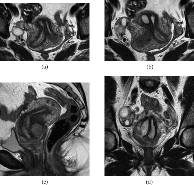
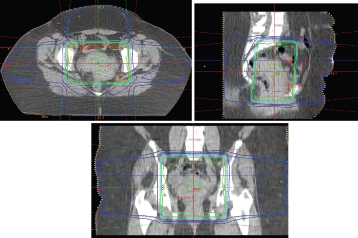
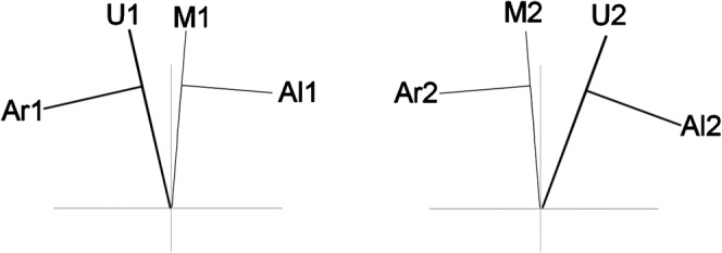
We report a case of congenital abnormality of bicornuate bicollis uterus in a patient who developed FIGO (International Federation of Gynecology and Obstetrics) stage IIB invasive carcinoma of the cervix in 2006. She was managed with radical concurrent chemoradiotherapy using an external photon beam of 50 Gy in 25 fractions and a weekly infusion of cisplatin, followed by low dose rate intracavity brachytherapy of 18 Gy to Manchester point A over two fractions. Intra-uterine afterloading brachytherapy catheters were inserted into both uterine cavities. Treatment was well tolerated with manageable acute toxicities. Complete response was achieved with therapy. The patient remains well on follow up with no clinical evidence of disease recurrence two years after initial treatment.
Figures
Similar articles
-
Bilateral radical radiotherapy in a patient with uterus didelphys.Br J Radiol. 2000 May;73(869):553-6. doi: 10.1259/bjr.73.869.10884756. Br J Radiol. 2000. PMID: 10884756
-
Concurrent chemoradiotherapy using high-dose-rate intracavitary brachytherapy for uterine cervical cancer.Gynecol Oncol. 2005 Mar;96(3):665-70. doi: 10.1016/j.ygyno.2004.11.046. Gynecol Oncol. 2005. PMID: 15721409
-
High complete response rate of concomitant chemoradiotherapy for locally advanced squamous cell carcinoma of the uterine cervix.Gynecol Oncol. 1996 Apr;61(1):101-8. doi: 10.1006/gyno.1996.0105. Gynecol Oncol. 1996. PMID: 8626094
-
Hyperfractionated radiotherapy with concurrent chemotherapy for para-aortic lymph node recurrence in carcinoma of the cervix.Int J Radiat Oncol Biol Phys. 2003 Apr 1;55(5):1247-53. doi: 10.1016/s0360-3016(02)04401-2. Int J Radiat Oncol Biol Phys. 2003. PMID: 12654434 Review.
-
Treatment results of high-dose-rate remote afterloading brachytherapy for cervical cancer and retrospective comparison of two regimens.Int J Radiat Oncol Biol Phys. 2003 Apr 1;55(5):1254-64. doi: 10.1016/s0360-3016(02)04525-x. Int J Radiat Oncol Biol Phys. 2003. PMID: 12654435 Review.
Cited by
-
Locally advanced adenocarcinoma of the cervix on uterus didelphys: a case report.J Contemp Brachytherapy. 2017 Feb;9(1):71-76. doi: 10.5114/jcb.2017.65640. Epub 2017 Jan 31. J Contemp Brachytherapy. 2017. PMID: 28344607 Free PMC article.
-
The Rare Condition of a Double Cervix: Results from the High-Risk Human Papillomavirus-Based Cervical Cancer Screening Program in the Lazio Region.Viruses. 2024 Jul 17;16(7):1149. doi: 10.3390/v16071149. Viruses. 2024. PMID: 39066311 Free PMC article.
-
The Utility of a Y-tandem Applicator in a Patient With Stage IVA Locally Advanced Cervical Cancer in the Setting of a Bicornuate Uterus.Cureus. 2025 Jan 8;17(1):e77151. doi: 10.7759/cureus.77151. eCollection 2025 Jan. Cureus. 2025. PMID: 39925542 Free PMC article.
-
Case report: Cervical brachytherapy technique for locally advanced cervical cancer in a patient with complete bicorporeal uterus.Front Oncol. 2024 Jun 5;14:1361562. doi: 10.3389/fonc.2024.1361562. eCollection 2024. Front Oncol. 2024. PMID: 38903713 Free PMC article.
-
Concurrent chemoradiotherapy for locally advanced squamous cell carcinoma of the cervix in a uterus didelphys with vaginal septum.J Contemp Brachytherapy. 2019 Apr;11(2):180-188. doi: 10.5114/jcb.2019.84506. Epub 2019 Apr 29. J Contemp Brachytherapy. 2019. PMID: 31139228 Free PMC article.
References
-
- Dean EM, Lambert GD, Dawes PJDK. Gynaecological treatments using the Selectron remote afterloading system. Br J Radiol 1988;61:1053–7 - PubMed
-
- Office forNationalStatistics. Cancer statistics registrations: registrations of cancer diagnosed in 2005, England. Series MB1 no.36. 2008.
-
- de Villiers EM, Fauquet C, Broker TR, Bernard HU, zur Hausen H. Classification of papillomaviruses. Virology 2004;324:17–27 - PubMed
-
- Wiley DJ, Douglas J, Beutner K, Cox T, Fife K, Moscicki AB, et al. External genital warts: diagnosis, treatment, and prevention. Clin Infect Dis 2002;35:5210–24 - PubMed
-
- Department ofHealth HPV vaccine recommended for NHS immunisation programme. London: DH, 2007
