. 2010 Sep 15;10(6):600-14.
doi: 10.4161/cbt.10.6.12586. Epub 2010 Sep 4.

ITF2 is a target of CXCR4 in MDA-MB-231 breast cancer cells and is associated with reduced survival in estrogen receptor-negative breast cancer

Affiliations

ITF2 is a target of CXCR4 in MDA-MB-231 breast cancer cells and is associated with reduced survival in estrogen receptor-negative breast cancer

Hitesh Appaiah et al. Cancer Biol Ther. .

Abstract

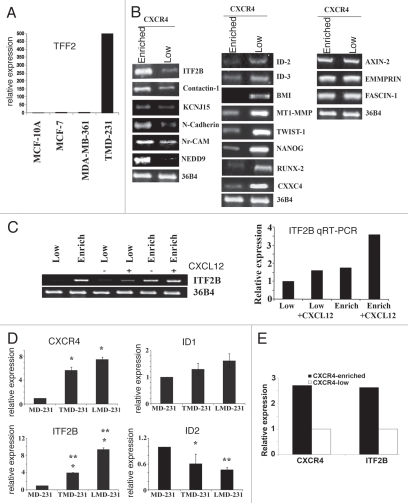
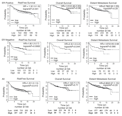
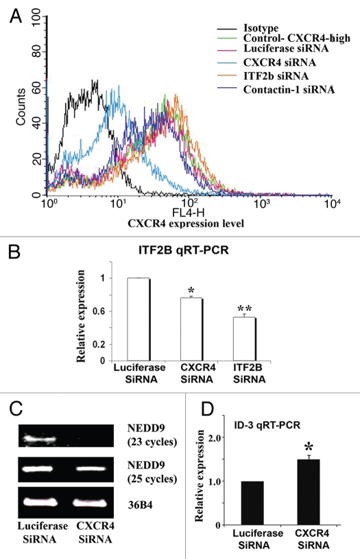
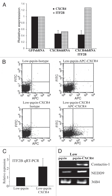
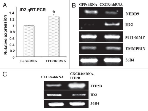
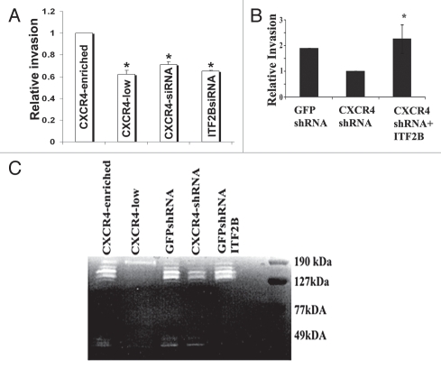
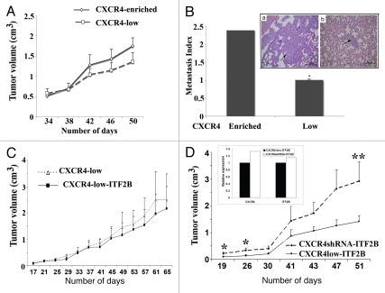
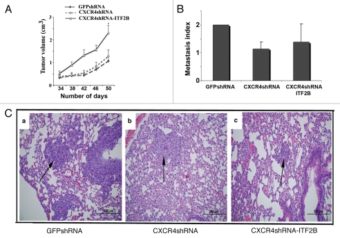
CXCR4, a chemokine receptor, plays an important role in breast cancer growth, invasion, and metastasis. The transcriptional targets of CXCR4 signaling are not known. Microarray analysis of CXCR4-enriched and CXCR4-low subpopulations of the MDA-MB-231 breast cancer cell line, which has a constitutively active CXCR4 signaling network, revealed differential expression of ∼ 200 genes in the CXCR4-enriched subpopulation. ITF2, upregulated in CXCR4-enriched cells, was investigated further. Expression array datasets of primary breast tumors revealed higher ITF2 expression in estrogen receptor negative tumors, which correlated with reduced progression free and overall survival and suggested its relevance in breast cancer progression. CXCL12, a CXCR4 ligand, increased ITF2 expression in MDA-MB-231 cells. ITF2 is a basic helix-loop-helix transcription factor that controls the epithelial-to-mesenchymal transition and the function of the ID family (inhibitor-of-differentiation) of transcription factors, such as ID2. ID2 promotes differentiation of breast epithelial cells and its reduced expression in breast cancer is associated with an unfavorable prognosis. Both CXCR4 and ITF2 repressed ID2 expression. In xenograft studies, CXCR4-enriched cells formed large tumors and exhibited significantly elevated lung metastasis. Short interfering RNA against ITF2 reduced invasion of the CXCR4-enriched MDA-MB-231 subpopulation, whereas ITF2 overexpression restored the invasive capacity of MDA-MB-231 cells expressing CXCR4shRNA. Furthermore, overexpression of ITF2 in these cells enhanced tumor growth. We propose that ITF2 is one of the CXCR4 targets, which is involved in CXCR4-dependent tumor growth and invasion of breast cancer cells.


Figures

Figure 1
Figure 1
CXCR4-dependent changes in gene expression in breast cancer cells. (A) TMD-231 cells express higher levels of TFF2, a CXCR4 ligand, compared to other breast cancer cell lines. TFF2 transcript levels were measured by qRT-PCR. (B) Semiquantitative RT-PCR analysis of differentially expressed genes in CXCR4-enriched and CXCR4-low subpopulation of TMD-231 cells. Genes expressed at higher (left), lower (middle) or similar levels (right) in CXCR4-enriched compared to CXCR4-low cells are shown. (C) The effect of CXCL12 on ITF2B expression in CXCR4-low and CXCR4-enriched cells. Cells were treated with CXCL12 (100 ng/ml for four hours) and ITF2B expression was measured by RT-PCR and qRT-PCR. 36B4 was used as a control for RT-PCR. (D) ITF2B expression is elevated in TMD-231 and LMD-231 cells compared to parental MDA-MB-231 (MD-231) cells. Note that ID2 expression is lower in TMD-231 and LMD-231 compared to parental MDA-MB-231 cells. For CXCR4, *p ≤ 0.006 MD-231 versus TMD-231 or LMD-231; for ITF2B, *p ≤ 0.0009 MD-231 versus TMD-231 or LMD-231; for ID2, *p = 0.02 MD-231 versus TMD-231, **p = 0.004 MD-231 versus LMD-231. Difference in expression of ITF2B was significantly different between TMD-231 and LMD-231 (**p = 0.002). (E) CXCR4 and ITF2B show similar pattern of expression in CXCR4-enriched and CXCR4-low cells. CXCR4 and ITF2B expression was measured by qRT-PCR.
Figure 2
Figure 2
ITF2B expression correlates with poor prognosis in breast cancer. Kaplan-Meier survival analyses of patients with low and high ITF2 (upper quartile) expressing tumors are shown. Data from patients who had not received any systemic therapy were included. Analyses were as follows: patients with ER-positive tumors (top), ER-negative (middle) and all patients (bottom). Number of patients at risk in each subgroups at 0, 5, 10, 15 and 20 years post diagnosis along with hazard ratio (HR) and log rank p values are indicated.
Figure 3
Figure 3
ITF2B is a downstream target of CXCR4. (A) Flow cytometry of TMD-231 treated with control luciferase siRNA, CXCR4 siRNA, ITF2B siRNA or Contactin-1 siRNA for CXCR4 expression. (B) CXCR4 siRNA treated cells show reduced ITF2B expression. ITF2B expression was measured by qRT-PCR. *p = 0.003, luciferase siRNA versus CXCR4; **p = 0.002 luciferase siRNA versus ITF2B siRNA. (C) CXCR4 siRNA treated cells show reduced NEDD9 expression. RT-PCR was used to measure NEDD9 expression. (D) CXCR4 siRNA treated cells show elevated ID3. qRT-PCR was used to measure ID3. *p = 0.02 luciferase siRNA versus CXCR4 siRNA.
Figure 4
Figure 4
The effects of CXCR4 knockdown and overexpression on ITF2B expression. (A) qRT-PCR showing CXCR4 and ITF2B expression in CXCR4-enriched cells stably expressing control GFPshRNA, CXCR4shRNA or CXCR4shRNA plus ITF2B. (B) Flow cytometry showing CXCR4 levels in CXCR4-low cells transduced with CXCR4 retrovirus. (C) CXCR4 overexpressing cells show elevated ITF2B. qRT-PCR was used to measure ITF2B expression. (D) CXCR4 overexpression also leads to elevated Contactin-1 and NEDD9.
Figure 5
Figure 5
ID2 is downstream of CXCR4 and ITF2B in TMD-231 cells. (A) Expression of ID2 in luciferase siRNA and ITF2BsiRNA treated cells. ID2 expression was measured by qRT-PCR. *p = 0.02, luciferase siRNA versus ITF2B siRNA treated cells. (B) CXCR4shRNA reduces NEDD9 but increases ID2 expression. (C) ITF2B suppresses ID2 expression in CXCR4shRNA cells.
Figure 6
Figure 6
ITF2B plays a role in invasion of CXCR4-enriched cells. (A) CXCR4siRNA and ITF2BsiRNA reduce the invasion of CXCR4-enriched cells in matrigel invasion assay (left part). *p = 0.02, CXCR4-enriched cells versus other cell types. (B) ITF2B overexpression restores invasive capacity to CXCR4-enriched cells expressing CXCR4shRNA. CXCL12 (100 ng/ml) was added to the bottom chamber in invasion assay. *p = 0.01 CXCR4 shRNA versus CXCR4shRNA-ITF2B. (C) CXCR4 and ITF2B control the expression of ∼130 kDa gelatinase. Conditioned media from indicated cell types were subjected to gelatin zymography. Representative gel from three experiments is shown.
Figure 7
Figure 7
Tumorigenic and metastatic properties of CXCR4-enriched and CXCR4-low cells. (A) Tumorigenic properties of CXCR4-enriched and CXCR4-low cells. Cells were injected into the mammary fat pad of female nude mice (n = 8 per group) and the tumor volume was measured on indicated days post injection. (B) Lung metastasis index in animals injected with CXCR4-enriched and CXCR4-low cells. H&E stained lung of a representative animal in both subgroups displaying a region of metastasis (indicated by an arrow) is shown. *p = 0.02, CXCR4-enriched versus CXCR4-low. (C) Tumorigenic properties of CXCR4-low cells with control vector or overexpressing ITF2B (n = 6). (D) Tumorigenic properties of CXCR4-low and CXCR4shRNA cells overexpressing ITF2B. These experiments were done in parallel (n = 6). *p = 0.03; **p = 0.07. Inset shows the expression levels of CXCR4 and ITF2B in two cell types.
Figure 8
Figure 8
Tumorigenic and metastasis properties of GFPshRNA, CXCR4shRNA and CXCR4shRNA-ITF2B cells. (A) Rate of tumor growth (n = 8 per group). *p = 0.01 (GFPshRNA or CXCR4shRNA cells versus CXCR4shRNA-ITF2B). (B) Lung metastasis index in GFPshRNA, CXCR4shRNA and CXCR4shRNA-ITF2B cell injected animals. (C) Representative lung metastasis in each group is shown.

Comment in

Similar articles

Cited by

References

    1. Eccles SA, Welch DR. Metastasis: recent discoveries and novel treatment strategies. Lancet. 2007;369:1742–1757. - PMC - PubMed
    1. Steeg PS. Tumor metastasis: mechanistic insights and clinical challenges. Nat Med. 2006;12:895–904. - PubMed
    1. Balkwill F. Cancer and the chemokine network. Nat Rev Cancer. 2004;4:540–550. - PubMed
    1. Orimo A, Gupta PB, Sgroi DC, Arenzana-Seisdedos F, Delaunay T, Naeem R, et al. Stromal fibroblasts present in invasive human breast carcinomas promote tumor growth and angiogenesis through elevated SDF-1/CXCL12 secretion. Cell. 2005;121:335–348. - PubMed
    1. Muller A, Homey B, Soto H, Ge N, Catron D, Buchanan ME, et al. Involvement of chemokine receptors in breast cancer metastasis. Nature. 2001;410:50–56. - PubMed

Publication types

MeSH terms

Substances

Associated data