Review
. 2010 Jul-Aug;7(4):441-52.
doi: 10.4161/rna.7.4.12258. Epub 2010 Jul 1.

Alternative splicing and muscular dystrophy

Affiliations
Review

Alternative splicing and muscular dystrophy

Mariaelena Pistoni et al. RNA Biol. 2010 Jul-Aug.

Abstract

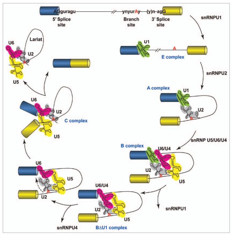
Alternative splicing of pre-mRNAs is a major contributor to proteomic diversity and to the control of gene expression in higher eukaryotic cells. For this reasons, alternative splicing is tightly regulated in different tissues and developmental stages and its disruption can lead to a wide range of human disorders. The aim of this review is to focus on the relevance of alternative splicing for muscle function and muscle disease. We begin by giving a brief overview of alternative splicing, muscle-specific gene expression and muscular dystrophy. Next, to illustrate these concepts we focus on two muscular dystrophy, myotonic muscular dystrophy and facioscapulohumeral muscular dystrophy, both associated to disruption of splicing regulation in muscle.


Figures

Figure 1
Figure 1
Schematic representation of the splicing reaction. Two exons (boxes) are separated by an intron (line). The consensus sequences in metazoans at the 5′ splice site, branch point and 3′ splice site are as indicated, where n is any nucleotide, r is a purine, and y is a pyrimidine. The polypyrimidine tract is a pyrimidine-rich stretch located between the branch site and the 3′ splice site. The cross-intron assembly and disassembly cycle of the major spliceosome is showed. The stepwise interaction of the spliceosomal snRNPs (colored structured RNAs) in the removal of the intron from the pre-mRNA is depicted.
Figure 2
Figure 2
Major forms of alternative splicing. Exons (boxes), introns (lines) alternative and splicing process (dotted lines). (A) Constitutive splicing. (B) Alternative use of an internal cassette exon. (C) Mutually exclusive exons. (D) Intron retention. (E) Alternative 5′ splice sites. (F) Alternative 3′ splice sites. (G) Alternative polyadenylation sites. (H) Alternative promoters. (I) Alternative last exons in which one exon contains the natural stop codon while the other exon contains a premature stop codon (PTC). In many cases, these common forms can be combined to generate more complicated alternative splicing events.
Figure 3
Figure 3
Examples of mutations causing splicing diseases and their possible consequences. Exons (boxes), introns (lines) and mutations (stars). (A–E) Cis-acting mutations on a specific pre-mRNA could lead to complete loss of a protein, the production of a mutant protein lacking a domain or to an unbalance in the abundance of the protein isoforms encoded by the pre-mRNA. (A) Mutations disrupting either 5′ or 3′ splice sites. (B) Mutations creating cryptic 5′ or 3′ splice sites. (C) Mutations disrupting splicing regulatory sequences such as intronic (ISS) or exonic (ESS) splicing silencers or intronic (ISE) or exonic (ESE) splicing enhancers. (D) Mutations creating new ISS, ESS, ISE or ESE. (E) Mutations affecting the secondary structure of the pre-mRNA. (F) Trans-acting mutations of a particular splicing regulator could lead to aberrant splicing of several different pre-mRNAs.
Figure 4
Figure 4
Model for the molecular consequences of triplet expansion in DM1. In healthy subjects, less than 40 CTG repeats are present in the 3′UTR of the DMPK gene and the DMPK and SIX5 genes are correctly expressed. In DM1 patients, CTG repeats are expanded to 80–1,000 repeats. This has a number of consequences. (A) Expanded repeats lead to epigenetic silencing of DMPK and SIX5 genes. (B) Long CUG repeats in the 3′UTR of the DMPK pre-mRNA titrate the RNA-binding protein MBNL1 leading to equivalent effects to MBNL1 loss-of-function. (C) Expanded CUG repeats cause phosphorylation and stabilization of the RNA-binding protein CUGBP1 leading to equivalent effects to CUGBP1 gain-of-function. (D) Altered activity/expression of MBNL1 and CUGBP1 is responsible for the alternative splicing changes observed in DM1. cTNT, IR and CLCN1 altered splicing is shown as example.
Figure 5
Figure 5
Model for the alternative splicing consequences of D4Z4 repeats deletion in FSHD. In healthy subjects, more than 10 D4Z4 repeats are present at the 4q35 sub-telomere and the FRG1 gene is correctly expressed. In FSHD patients, D4Z4 repeats are deleted leaving 1-10. This leads to the epigenetic overexpression of FRG1. Increased FRG1 expression is responsible for the alternative splicing changes observed in FSHD.

Similar articles

Cited by

References

    1. Wahl MC, Will CL, Luhrmann R. The spliceosome: design principles of a dynamic RNP machine. Cell. 2009;136:701–18. - PubMed
    1. Wang GS, Cooper TA. Splicing in disease: disruption of the splicing code and the decoding machinery. Nat Rev Genet. 2007;8:749–61. - PubMed
    1. Wang Z, Burge CB. Splicing regulation: from a parts list of regulatory elements to an integrated splicing code. RNA. 2008;14:802–13. - PMC - PubMed
    1. Fu XD. Towards a splicing code. Cell. 2004;119:736–8. - PubMed
    1. Zhang XH, Chasin LA. Computational definition of sequence motifs governing constitutive exon splicing. Genes Dev. 2004;18:1241–50. - PMC - PubMed

Publication types

Substances