Review
. 2010:26:667-87.
doi: 10.1146/annurev-cellbio-100109-103948.

Stem cell models of cardiac development and disease

Affiliations
Review

Stem cell models of cardiac development and disease

Kiran Musunuru et al. Annu Rev Cell Dev Biol. 2010.

Abstract

The past few years have witnessed remarkable advances in stem cell biology and human genetics, and we have arrived at an era in which patient-specific cell and tissue models are now practical. The recent identification of cardiovascular progenitor cells, as well as the identification of genetic variants underlying congenital heart disorders and adult disease, opens the door to the development of human models of human cardiovascular disease. We review the current understanding of the contribution of progenitor cells to cardiogenesis and outline how pluripotent stem cells can be applied to the modeling of cardiovascular disorders of genetic origin. A key challenge will be to implement these models in an efficient manner to develop a molecular understanding of how genes lead to disease and to screen for genes and drugs that modify the disease process.


Figures

Figure 1
Figure 1
Embryological origin of the mammalian heart. Early in cardiac development, cardiac progenitors from the first heart field (FHF) coalesce to form the cardiac crescent. Progenitors from the second heart field (SHF) localize anterior and medial to cells of the FHF. As development progresses, the heart tube forms and then loops to ultimately form the primitive chambers of the four chambered mammalian heart. Progenitors from the FHF most greatly contribute to the left ventricle, whereas progenitors from the SHF cells contribute the majority of the cells of the right ventricle and outflow tract. Progenitors from both heart fields contribute to the inflow tract. In mature heart, SHF cells contribute to the majority of the cells of the heart except for the left ventricular free wall., FHF cells contribute to the left ventricle, the ventricular septum, and both atria. Green = areas with FHF cells; red = areas with SHF cells; brown = areas with both FHF and SHF cells. Ao = aorta; CC = cardiac crescent; OT = outflow tract; IT = inflow tract; LA = left atrium; LV = left ventricle; HF = head fold; ML = midline; PA = pulmonary artery; RA = right atrium; RV = right ventricle.
Figure 2
Figure 2
Proposed cell fate map for progenitor cells during development. Following gastrulation, multipotent cardiac progenitor cells responsible for populating the FHF and SHF emerge from cardiac mesoderm. Multipotent Isl1+ progenitors from the SHF give rise to endothelium, smooth muscle, and cardiac muscle; Isl– progenitors appear to give rise to at least smooth muscle and cardiac muscle.
Figure 3
Figure 3
Strategies for obtaining disease-specific human cardiomyocytes and other cell types. Disease-specific human embryonic stem (ES) cells can be derived via homologous recombination in normal ES cells to insert disease-associated mutations. Alternatively, patient-specific induced pluripotent stem (iPS) cells can be derived directly from patient-specific somatic cells (typically skin fibroblasts). Either type of disease-specific pluripotent stem cells can be differentiated into various cell types, e.g., into multipotent cardiac progenitors that can be further differentiated into cardiomyocytes, smooth muscle cells, or endothelial cells.
Figure 4
Figure 4
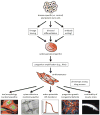
Scheme for the derivation of disease-specific cardiomyocytes for use in phenotypic assays and drug screens. Disease-specific pluripotent stem cells will undergo in vitro differentiation and cardiac progenitors will be purified (via any of several techniques, including lineage tracing, directed differentiation, and antibody sorting). Purified populations of cardiac progenitors will then be expanded into larger numbers, followed by further differentiation into functional cardiomyocytes. Cardiomyocytes will then be used in various cellular assays, several examples of which are shown, to study and develop therapies for a variety of cardiovascular disorders, including cardiomyopathy, electrophysiological disorders, and congenital defects.

References

    1. Agarwal S, Holton KL, Lanza R. Efficient differentiation of functional hepatocytes from human embryonic stem cells. Stem Cells. 2008;26:1117–27. - PubMed
    1. Amit M, Carpenter MK, Inokuma MS, Chiu CP, Harris CP, et al. Clonally derived human embryonic stem cell lines maintain pluripotency and proliferative potential for prolonged periods of culture. Dev Biol. 2000;227:271–8. - PubMed
    1. Basson CT, Bachinsky DR, Lin RC, Levi T, Elkins JA, et al. Mutations in human TBX5 cause limb and cardiac malformation in Holt-Oram syndrome. Nat Genet. 1997;15:30–5. - PubMed
    1. Benson DW, Silberbach GM, Kavanaugh-McHugh A, Cottrill C, Zhang Y, et al. Mutations in the cardiac transcription factor NKX2.5 affect diverse cardiac developmental pathways. J Clin Invest. 1999;104:1567–73. - PMC - PubMed
    1. Bruneau BG. The developmental genetics of congenital heart disease. Nature. 2008;451:943–8. - PubMed

Substances