Review
. 2010:26:179-210.
doi: 10.1146/annurev-cellbio-100109-104129.

Ubiquitination in postsynaptic function and plasticity

Affiliations
Review

Ubiquitination in postsynaptic function and plasticity

Angela M Mabb et al. Annu Rev Cell Dev Biol. 2010.

Abstract

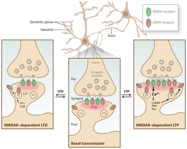
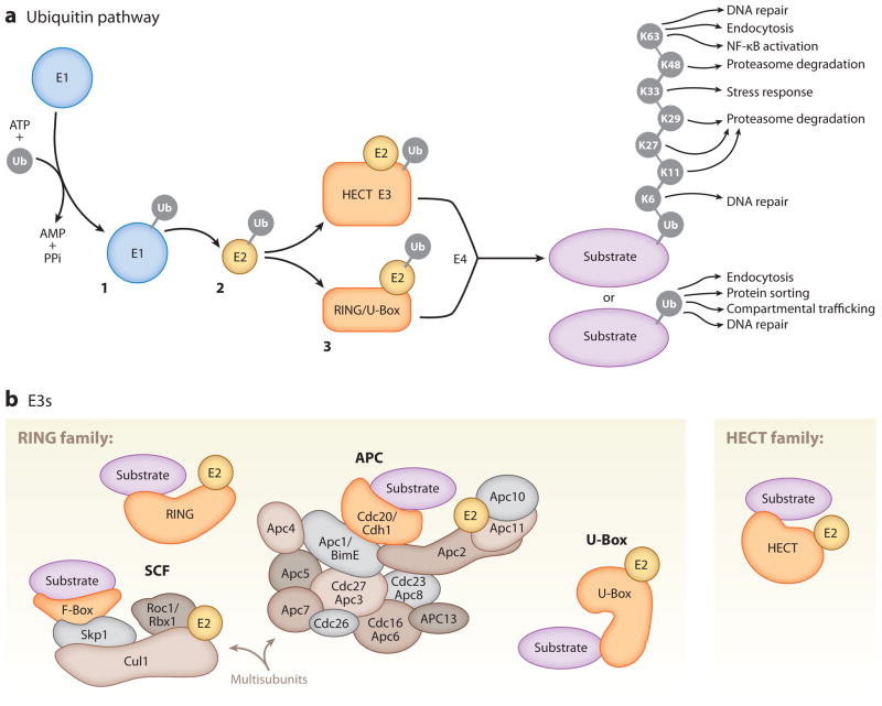
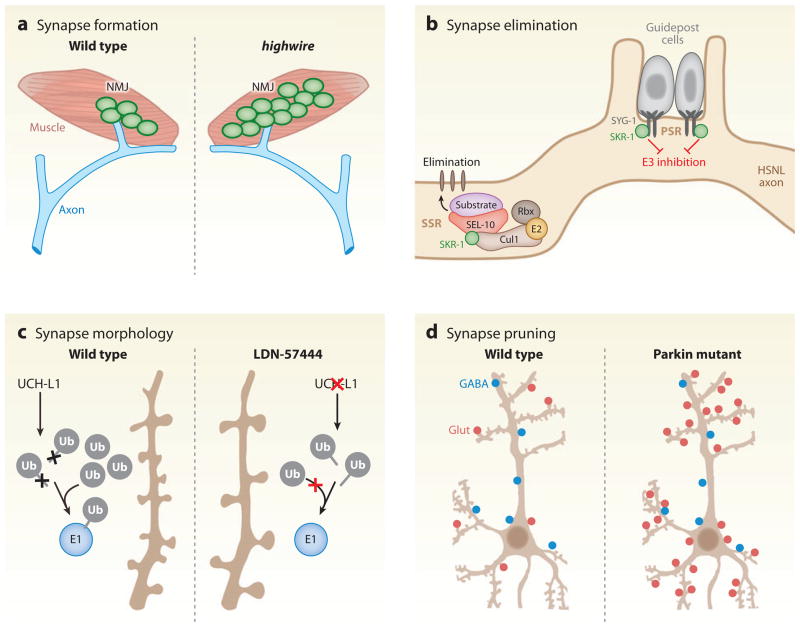
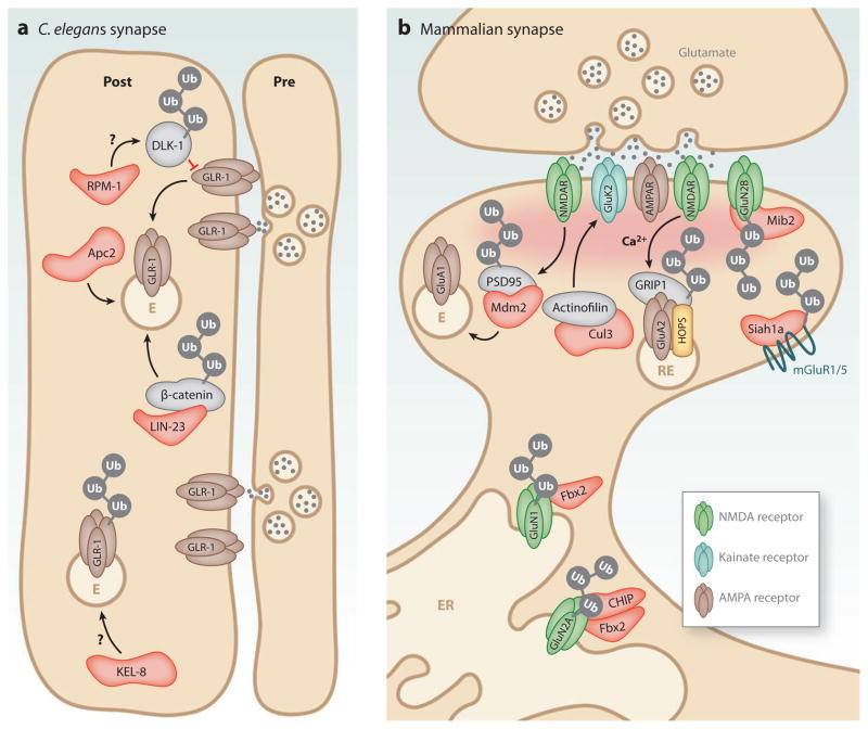
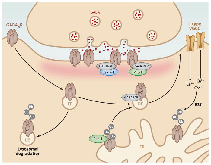
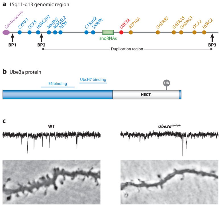
Neurons are highly specialized cells whose connectivity at synapses subserves rapid information transfer in the brain. Proper information processing, learning, and memory storage in the brain requires continuous remodeling of synaptic networks. Such remodeling includes synapse formation, elimination, synaptic protein turnover, and changes in synaptic transmission. An emergent mechanism for regulating synapse function is posttranslational modification through the ubiquitin pathway at the postsynaptic membrane. Here, we discuss recent findings implicating ubiquitination and protein degradation in postsynaptic function and plasticity. We describe postsynaptic ubiquitination pathways and their role in brain development, neuronal physiology, and brain disorders.


Figures

Figure 1
Figure 1
Bidirectional plasticity at excitatory synapses. Typical synapses in the central nervous system consist of an axon serving as the presynaptic terminal in a junctional contact with the postsynaptic cell. The postsynaptic membrane contains a dense matrix organized in an array of scaffolding molecules known as the postsynaptic density (PSD, pink), which allows for the anchoring and positioning of receptors, such as α-amino-3-hydroxyl-5-methyl-4-isoxazole propionic acid (AMPA) receptors and N-methyl-D-aspartic acid (NMDA) receptors (NMDARs), for efficient activation by presynaptically released glutamate neurotransmitter (middle inset). Learning-related brain plasticity involves the strengthening or weakening of synapses. At excitatory glutamatergic synapses, such synaptic plasticity can be finely tuned by altering the channel properties or abundance of glutamate receptors within the PSD. A prolonged enhancement of synaptic transmission at a synapse is known as long-term potentiation (LTP), whereas a persistent weakening of synaptic transmission at a synapse is known as long-term depression (LTD) (Malenka & Bear 2004). LTP is the most celebrated cellular model for learning and memory in the brain and can be subdivided into two temporal phases that differ in their mechanism of expression. The initial phase of NMDAR-dependent LTP, termed early phase LTP (E-LTP), lasts for ~1–2 hours. E-LTP requires Ca2+ influx through NMDARs and activation of CaMKII, and it is expressed in part by an increase in the number of AMPA receptors trafficked through recycling endosomes (RE). The second phase, known as late phase LTP (L-LTP), requires protein synthesis and gene transcription in the postsynaptic cell to allow for long-lasting enhancement of synaptic transmission for several hours to weeks (Malenka & Bear 2004). Structural remodeling such as increases in PSD size, the growth of new dendritic spines, and the enlargement of preexisting spines is also associated with LTP (Holtmaat & Svoboda 2009). Several forms of LTD contribute to learning-related plasticity at glutamatergic synapses. Various forms of LTD require NMDARs, group I metabotropic glutamate receptors (mGluRs), and endocannabinoid signaling (Malenka & Bear 2004). NMDAR-dependent LTD in CA1 hippocampus is the best-studied form of LTD and, like LTP, its induction requires Ca2+ influx through NMDARs, but it depends on protein phosphatase 1 (PP1) and calcineurin (CaN). The expression of mGluR-LTD requires protein synthesis. In general, LTD is accompanied by a reduction in the number of AMPA receptors that are removed by endocytosis and transported to early endosomes (EE) (Malenka & Bear 2004).
Figure 2
Figure 2
Components of the ubiquitin (Ub) pathway. (a) Substrate ubiquitination requires a three-step enzymatic reaction. In step 1, a ubiquitin-activating enzyme (E1) activates free ubiquitin, a process in which the C-terminal glycine residue of ubiquitin is coupled to a cysteine residue of the E1 via a thioester linkage. In step 2, the activated ubiquitin is transferred to a cysteine residue of a ubiquitin-conjugating enzyme (E2) and linked via a thioester linkage. In step 3, a ubiquitin ligase (E3) transfers ubiquitin to the substrate via an amide isopeptide linkage to the ε-amino group of the substrate lysine (Hershko & Ciechanover 1998). In some cases, a polyubiquitin ligase (E4) coordinates with a ubiquitin E3 to lengthen ubiquitin chains (Hoppe 2005). Ubiquitin can be attached to lysine residues of previously conjugated ubiquitins in multiple polyubiquitin chain configurations (K6, K11, K27, K29, K33, K48, K63) to regulate various functions. (b) Ubiquitin E3s are divided into the RING (really interesting new gene) and HECT (homologous to E6-associated protein C terminus) domain families, among others. The RING domain family is the largest family of ubiquitin E3s. RING ubiquitin E3s function as single subunits or in a multisubunit complex. Illustrated here are the best-studied multisubunit RING ubiquitin E3s, the Skp1/Cullin/F-box (SCF) and anaphase-promoting complex/cyclosome (APC/C) complexes. U-Box proteins are a distinct set of RING-like ubiquitin E3s.
Figure 3
Figure 3
Ubiquitination in synapse regulation. (a) Loss of function of the ubiquitin E3 highwire (hiw) leads to overgrowth of presynaptic terminals at the Drosophila neuromuscular junction (NMJ) (DiAntonio et al. 2001). (b) Sequestration and inhibition of the SCF ubiquitin E3 subunit, SKR-1, by its binding to the synaptic adhesion molecule SYG-1 prevents synapse elimination in the Caenorhabditis elegans hermaphrodite-specific motor neuron (HSNL) axon at the primary synapse region (PSR). During development, synapses in the HSNL axon are eliminated in the secondary synapse region (SSR) owing to the lack of SKR-1 inhibition and corresponding assembly of a functional SKR-1 SCF ubiquitin E3 complex composed of SKR-1, CUL1, SEL-10, and Rbx (Ding et al. 2007). (c) In hippocampal cultures, inhibition of the DUB UCH-L1 by the isatin O-acyl oxime compound, LDN-57444, decreases spine density and increases spine length and width, likely by depletion of free ubiquitin (Cartier et al. 2009). (d) In hippocampal cultures, loss of the RING E3 ligase parkin or expression of Parkinson’s disease–linked mutant forms of parkin increases the number of excitatory synapses (Glut, red circles) with no change in inhibitory synapse number (GABA, blue circles) (Helton et al. 2008).
Figure 4
Figure 4
Postsynaptic control of glutamate receptors by ubiquitin E3 ligases. Ubiquitin E3 ligase subunits that have been implicated in glutamate receptor regulation are indicated in red. (a) At C. elegans synapses, ubiquitination of the MAPKKK DLK-1 by SCFRPM-10, ubiquitin-dependent degradation of β-catenin by LIN-23 (C. elegans ortholog of βTrCP), and APC/C ubiquitin E3 activity (Apc2) all lead to a loss of GLR-1-containing AMPA receptors through the endocytic pathway. Ubiquitination by the KEL-8/Cul3 cullin complex also leads to loss of GLR-1 at synapses. E, endosome. (b) At mammalian synapses, Mdm2-dependent ubiquitination of PSD-95 leads to a loss of GluA1-containing AMPA receptors (AMPARs), likely through the endocytic pathway. The ubiquitin-like domain protein Tmub1/HOPS (transmembrane and ubiquitin-like domain-containing 1/Hepatocyte Odd Protein Shuttling) is associated with recycling endosomes (REs) and promotes GluA2 AMPAR recycling in coordination with GRIP1 (glutamate receptor interacting protein 1). The glycosylated NMDA receptor (NMDAR) subunit, GluN1, is ubiquitinated by SCFFbx2 in the endoplasmic reticulum (ER) and is required for surface expression of NMDARs. The NMDAR subunit GluN2A is ubiquitinated by the co-chaperone/ubiquitin ligase C terminus of Hsc-70-interaction protein (CHIP) and SCFFbx2 while in the ER. Ubiquitination of GluN2B by the RING ubiquitin E3 mindbomb2 (Mib2) leads to a loss of GluN2B-containing receptors from the synapse. The ubiquitin RING E3 ligase Siah1a (seven in absentia homolog) is required for ubiquitination of mGluR1 and mGluR5, which leads to their removal from the plasma membrane.
Figure 5
Figure 5
Ubiquitination at inhibitory synapses. At inhibitory synapses, ubiquitination of the γ2 subunit of GABAA receptors (GABAARs) results in lysosomal degradation of GABAARs. Binding of Plic-1 to GABAARs in the endoplasmic reticulum (ER) leads to stabilization of ubiquitinated GABAARs and promotes their forward trafficking. Interactions of GABAAR with the ubiquitin-like proteins GABARAP and Plic-1 promote recycling and aid in postsynaptic clustering of GABAARs. Interactions of GABARAP with GRIP-1 further aid in the clustering of postsynaptically expressed GABAARs. Ubiquitination and destabilization of GABAARs in the ER are accelerated by Ca2+ signaling through L-type voltage-gated calcium channels (VGCCs) by an unknown ubiquitin E3 ligase. EE, early endosome; LE, late endosome; RE, recycling endosome.
Figure 6
Figure 6
The HECT domain E3 ligase Ube3a regulates the number and function of glutamatergic synapses. (a) Location of the UBE3A gene within the human chromosome region 15q11-q13 that is linked to autism. Maternally inherited deletions within the 15q11-q13 region and mutations within Ube3a cause Angelman syndrome, whereas duplications within the 15q11-q13 region are associated with autism. Paternally expressed genes are depicted in blue, whereas maternally expressed genes are depicted in red. Nonimprinted genes are represented in gold. Common breakpoint regions (BP1–3) are represented by the vertical black arrows. Class I deletions (BP1–BP3) and class II deletions (BP2 and BP3) are both associated with Prader-Willi syndrome and Angelman syndrome. (b) Domain structure of the Ube3a protein. Ube3a contains a HECT domain (gray) that is required for the ubiquitin ligase activity of Ube3a. Ubiquitin is conjugated to cysteine 833 within the HECT domain and is required for the ubiquitin E3 activity of Ube3a. Interacting regions for the E2 UbcH7 and the viral oncogenic protein E6 are indicated. (c) Loss of Ube3a impairs synapse development. Traces indicate miniature excitatory postsynaptic currents (mEPSCs) recorded from layer 2/3 pyramidal neurons in visual cortex slices from wild-type (WT) or Ube3am−/p+ juvenile (~P25) mice showing a reduction in mEPSC frequency in Ube3am−/p+ neurons. Lower panels indicate basal dendrites in layer 2/3 pyramidal neurons showing a reduction in dendritic spines in Ube3am−/p+ mice. Figure adapted with permission from Yashiro et al. (2009).

References

    1. Ahmari SE, Buchanan J, Smith SJ. Assembly of presynaptic active zones from cytoplasmic transport packets. Nat Neurosci. 2000;3:445–51. - PubMed
    1. Anderson C, Crimmins S, Wilson JA, Korbel GA, Ploegh HL, Wilson SM. Loss of Usp14 results in reduced levels of ubiquitin in ataxia mice. J Neurochem. 2005;95:724–31. - PubMed
    1. Ang XL, Seeburg DP, Sheng M, Harper JW. Regulation of postsynaptic RapGAP SPAR by Polo-like kinase 2 and the SCFβ-TRCP ubiquitin ligase in hippocampal neurons. J Biol Chem. 2008;283:29424–32. - PMC - PubMed
    1. Arancibia-Carcamo IL, Yuen EY, Muir J, Lumb MJ, Michels G, et al. Ubiquitin-dependent lysosomal targeting of GABAA receptors regulates neuronal inhibition. Proc Natl Acad Sci USA. 2009;106:17552–57. - PMC - PubMed
    1. Artinian J, McGauran AM, De Jaeger X, Mouledous L, Frances B, Roullet P. Protein degradation, as with protein synthesis, is required during not only long-term spatial memory consolidation but also reconsolidation. Eur J Neurosci. 2008;27:3009–19. - PubMed

Publication types

Substances

LinkOut - more resources